. 2015 Dec;1(1):6.
doi: 10.1186/s40792-014-0013-1. Epub 2015 Jan 28.

Primary intrathoracic malignant neurogenic tumor: report of three cases and comparison with benign neurogenic tumors resected at our institution

Affiliations

Primary intrathoracic malignant neurogenic tumor: report of three cases and comparison with benign neurogenic tumors resected at our institution

Takeshi Kawaguchi et al. Surg Case Rep. 2015 Dec.

Abstract

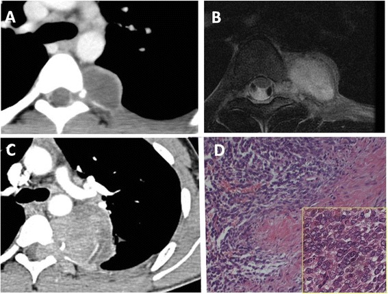
We present three patients with intrathoracic malignant neurogenic tumor. Two lesions showed no sign of invasion into adjacent structures, while the third lesion extended to the intraspinal canal with vertebral involvement. Although all three lesions were completely excised, each patient relapsed within 1 year of the initial treatment. One patient with local recurrence underwent radiation therapy, but the recurrent tumor continued to progress. Chemotherapy was subsequently performed. Two patients with distant metastases also received chemotherapy. Because there is no effective chemotherapeutic regimen for intrathoracic malignant neurogenic tumor, all three patients received high-dose chemotherapy followed by hematopoietic stem cell transplantation. Although the relapsed lesions temporarily regressed after treatment, all three patients showed disease recrudescence and ultimately died of their disease. A comparison of the intrathoracic malignant neurogenic tumors and the benign neurogenic tumors resected at our institution revealed no meaningful differences distinguishing malignant from benign neurogenic tumors prior to surgery.

Keywords: Malignant tumor; Mediastinal tumor; Neurogenic tumor; Prognosis; Surgery.


Figures

Figure 1
Figure 1
Diagnostic exam results for case 1. Chest enhanced CT (A) and T2-weighted MRI (B) showed a well-defined and ovoid mass located in the paravertebral sulcus without invasion of the vertebral body or intraspinal canal. The microscopic appearance of the area with a solid growth of primitive cells is shown (C). The lesion was highly cellular (low-power view). The tumor nest was composed of primitive cells with round or oval hyperchromatic nuclei and scant cytoplasm (high-power view).
Figure 2
Figure 2
Diagnostic exam results for case 2. Chest unenhanced CT (A) and T2-weighted MRI (B) showed a well-defined and round mass located in the paravertebral sulcus without invasion of the vertebral body or intraspinal canal. The microscopic appearance of the tumor is shown (C). It was a hypercellular, fascicular spindle cell neoplasm; the cells within the lesion had tapering nuclei and pale, indistinct cytoplasm (low-power view). Within these overtly malignant areas, numerous large, bright eosinophilic rhabdomyoblasts were seen (high-power view).
Figure 3
Figure 3
Diagnostic exam results for case 3. Chest enhanced CT (A) and T2-weighted MRI (B) showed a well-defined and ovoid mass located in the paravertebral sulcus. The tumor extended into the spinal canal. Six months after initial detection of the tumor, chest enhanced CT (C) showed a huge, heterogeneous mass extending into the intraspinal canal with involvement of adjacent vertebrae. The microscopic appearance of the tumor is shown (D). The tumor was a highly cellular malignant spindle cell neoplasm that contained alternating cellular and more myxoid areas (low-power view). The tumor cells had eosinophilic cytoplasm, wavy hyperchromatic nuclei, and high mitotic activity (high-power view).

References

    1. Strollo DC, Rosado-de-Christenson ML, Jett JR. Primary mediastinal tumors: part II. Tumors of the middle and posterior mediastinum. Chest. 1997;112:1344–57. doi: 10.1378/chest.112.5.1344. - DOI - PubMed
    1. Shields TW. Benign and malignant neurogenic tumors of the mediastinum in adults. In: Shields TW, LoCicero J III, Ponn RB, editors. General thoracic surgery. Philadelphia: Lippincott Williams & Wilkins; 2000. pp. 2313–27.
    1. Kourea HP, Bilsky MH, Leung DHY, Lewis JJ, Woodruff JM. Subdiaphragmatic and intrathoracic paraspinal malignant peripheral nerve sheath tumors. Cancer. 1998;82:2191–203. doi: 10.1002/(SICI)1097-0142(19980601)82:11<2191::AID-CNCR14>3.0.CO;2-P. - DOI - PubMed
    1. Aydm GB, Kutluk MT, Yalçin B, Büyükpamukçu M, Kale G, Varan A, et al. Neuroblastoma in Turkish children. J Pediatr Hematol Oncol. 2009;31:471–80. doi: 10.1097/MPH.0b013e3181a6dea4. - DOI - PubMed
    1. Stucky CC, Johnson KN, Gray RJ, Pockaj BA, Ocal IT, Rose PS, et al. Malignant peripheral nerve sheath tumors (MPNST): the Mayo Clinic experience. Ann Surg Oncol. 2012;19:878–85. doi: 10.1245/s10434-011-1978-7. - DOI - PubMed

LinkOut - more resources