. 2015 Dec;1(1):9.
doi: 10.1186/s40792-014-0007-z. Epub 2015 Jan 30.

A case of mucinous cystic neoplasm of the liver: a case report

Affiliations

A case of mucinous cystic neoplasm of the liver: a case report

Yusuke Nakayama et al. Surg Case Rep. 2015 Dec.

Abstract

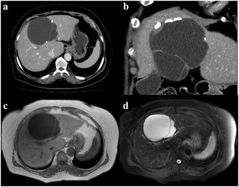
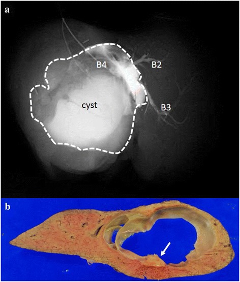
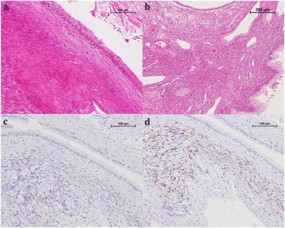
A 71-year-old woman was referred to our institution for further investigation of epigastric pain. The patient had been detected to have a multilocular cyst in the medial segment of the liver measuring 69 mm in diameter at another hospital 2 years ago, and the diameter of the cyst had increased to 90 mm. Although the cyst had gradually increased in size, there was no evidence of mural nodules. As we were concerned about the malignant potential of the lesion, a left hepatic segmentectomy was performed. Pathologically, the cyst was lined by columnar and cuboidal epithelium with low-grade atypia. The epithelium covered an ovarian-like stroma, and the diagnosis was mucinous cystic neoplasm of the liver (MCN-L) with low-grade intraepithelial neoplasia. MCN-L is a rare disease and its characteristics are still poorly understood. MCN-L occurs at a lower frequency as compared to the counterpart of MCN of the pancreas, further investigations are necessary to clarify the biological malignancy of MCN-L.

Keywords: Mucinous cystic neoplasm of the liver; Ovarian-like stroma; Prognosis.


Figures

Figure 1
Figure 1
CT and T1- and T2-weighted images. CT showing a multilocular cystic lesion in the medial segment of the liver with calcification in the cyst wall (a). Thickening of the cyst wall around the calcification (b). Contrast-enhanced CT showed no enhancement of the thickened wall (a, b). The cystic lesion was visualized as a hypointensity on T1-weighted images (c) and as a hyperintensity on the T2-weighted images (d).
Figure 2
Figure 2
The cyst showed contrast enhancement following postoperative injection of contrast medium into B4. The dotted line shows an outline of the cyst (a). The resected specimen was a multilocular cystic lesion covered by a thick fibrous capsule (b). Calcification was found in the cyst wall (arrow). No mural nodules were found in the cystic lesion.
Figure 3
Figure 3
The cyst and stromal cells. The cyst was lined by cuboidal columnar epithelium, with low-grade atypia of the epithelial cells ((a) H&E staining, ×200). The columnar and cuboidal epithelium covered an ovarian-like hypercellular stroma ((b) H&E staining, ×100). This stroma was immunoreactive for estrogen and progesterone receptors ((c) estrogen receptor staining, ×200; (d) progesterone receptor staining, ×200).

References

    1. Tsui WMS, Adsay NV, Crawford JM, Hruban R, Kloppel G, Wee A, editors. WHO classification of tumors of the digestive system. 4. Lyon: WHO; 2010. pp. 236–8.
    1. Simo KA, Mckillop LH, Ahrens WA, Martinie JB, Lannitti DA, Sindram D. Invasive biliary mucinous cystic neoplasm: a review. HPB. 2012;14:725–40. doi: 10.1111/j.1477-2574.2012.00532.x. - DOI - PMC - PubMed
    1. Kubota K, Nakanuma Y, Kondo F, Hachiya H, Miyazaki M, Nagino M, et al. Clinicopathological features and prognosis of mucin-producing bile duct tumor and mucinous cystic tumor of the liver: a multi-institutional study by the Japan Biliary Association. J Hepatobiliary Pancreat Sci. 2014;21:176–85. doi: 10.1002/jhbp.23. - DOI - PubMed
    1. Shiono S, Suda K, Nobukawa B, Arakawa A, Yamasaki S, Sasahara N, et al. Pancreatic, hepatic, splenic, and mesenteric mucinous cystic neoplasms (MCN) are lumped together as extraovarian MCN. Pathol Int. 2006;56:71–7. doi: 10.1111/j.1440-1827.2006.01926.x. - DOI - PubMed
    1. Lim JH, Zen Y, Jang KT, Kim YK, Nakanuma Y. Cyst-forming intraductal papillary neoplasm of the bile ducts: description of imaging and pathologic aspects. AJR. 2011;197:1111–20. doi: 10.2214/AJR.10.6363. - DOI - PubMed

LinkOut - more resources