

Primary leiomyosarcoma of the pancreas: report of a case treated by local excision and review of the literature
- PMID: 26943422
- PMCID: PMC4595416
- DOI: 10.1186/s40792-015-0097-2
Primary leiomyosarcoma of the pancreas: report of a case treated by local excision and review of the literature
Abstract
Background: First described by Ross in 1951, primary pancreatic leiomyosarcoma is a rare mesenchymal tumour of the pancreas, with nonspecific clinical and radiological features and a poor prognosis, if unresectable.
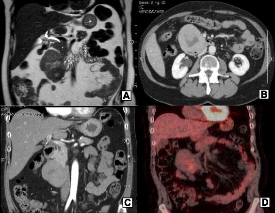
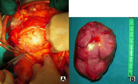
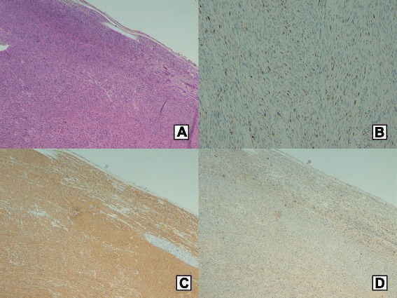
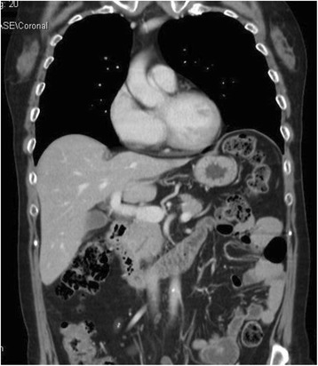
Case report: A 60-year-old woman presented with abdominal pain. Magnetic resonance imaging (MRI) and computed tomography (CT) scan detected a dishomogeneous egg-shaped 8-cm mass, arising from the pancreatic head, with duodenal compression, without dilation of the Wirsung duct. (18)F-FDG positron-emission tomography (PET)-CT showed a moderate tracer uptake, and the endoscopic ultrasound (US) showed a hypoechoic lesion, arising from the duodenal wall, suspected to be a gastrointestinal stromal tumour (GIST). CEA, CA19-9, NSE, and chromogranin A were normal. At the surgical exploration, a 10-cm mass, adherent to the anterior aspect of the pancreatic head, was found. The lesion was easily separable from the duodenal wall and was totally excised. The frozen intraoperative examination showed a mesenchymal tumour, with spindle-shaped cells, suggesting that a GIST diagnosis was likely. Postoperative course was uneventful. Histology and immunohistochemistry demonstrated a well-differentiated leiomyosarcoma, with five to six mitotic counts per 10 high-power field (HPF) and proliferative index (MIB-1) 10 % (grade 2 according to Federation Nationale des Centres de Lutte Contre le Cancer (FNCLCC)), with positive smooth muscle actin, desmin, and caldesmon but negative CD117 (c-kit) and S-100. The patient is alive and asymptomatic 19 months after surgery, without evidences of disease.
Conclusions: In the English literature, only 44 cases of primary pancreatic leiomyosarcoma have been reported. If a pancreatic mass suspected for primary pancreatic leiomyosarcoma has no adjacent organ/vessel invasion or distant metastases, surgical resection is the therapy of choice.
Keywords: Leiomyosarcoma; Mesenchymal tumour; Pancreas; Pancreatic tumour; Sarcoma.
Figures
References
-
- Klimstra DS, Hruban RH, Pitman MB. Pancreas. In: Mills SE, editor. Histology for pathologists. 4. Philadelphia, PA: Lippincott Williams & Wilkins; 2012. pp. 777–816.
-
- Miettinen M, Fletcher CDM, Kindblom LG, Tsui WMS. Mesenchymal tumors of the pancreas. In: Bosman FT et al., editors. WHO classification of tumours of the digestive system. 4th ed. Lyon, France: International Agency for Research on Cancer; 2010. p. 331.
LinkOut - more resources
Full Text Sources
Other Literature Sources
Research Materials
