. 2015 Dec;1(1):107.
doi: 10.1186/s40792-015-0110-9. Epub 2015 Oct 17.

Pseudolymphoma of the liver: a case report and literature review

Affiliations

Pseudolymphoma of the liver: a case report and literature review

Kazuhiro Taguchi et al. Surg Case Rep. 2015 Dec.

Abstract

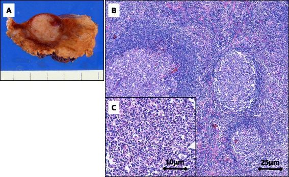
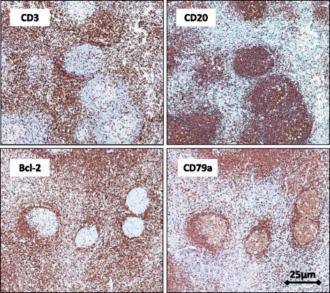
Pseudolymphoma is a benign lymphocytic tumor-like lesion, and its occurrence in the liver is rare. Here, we report the case of a 78-year-old woman with pseudolymphoma of the liver. She had a history of tremors for several years. Therefore, she underwent computed tomography (CT) for screening, and liver tumors were incidentally identified. She did not have any history of liver disease. Liver function test results and tumor marker levels were all within normal limits, and viral markers for hepatitis were negative. Contrast-enhanced CT revealed four nodules measuring up to 13 mm in diameter with ring enhancement in both lobes of the liver. On magnetic resonance imaging, the lesions showed slightly high intensity on T2-weighted images and high intensity on diffusion-weighted images. Because of atypical imaging findings, the tumors could not be definitively diagnosed. Therefore, we performed laparoscopic limited resection of segments 2, 3, 4, and 8 of the liver. The final pathological diagnosis was pseudolymphoma of the liver. The patient has had no signs of recurrence for 6 months after the surgery. Although pseudolymphoma of the liver is rare, it is necessary to consider it in the differential diagnosis of a liver tumor.

Keywords: Laparoscopic hepatectomy; Liver; Pseudolymphoma.


Figures

Fig. 1
Fig. 1
Contrast-enhanced computed tomography showing a nodule with ring enhancement in the arterial phase and subsequent washout in the portal phase in segments 2, 3 (arrow), 4, and 8 of the liver
Fig. 2
Fig. 2
Magnetic resonance imaging (MRI) showing a nodule with slight high intensity on a T2-weighted image and high intensity on a diffusion-weighted image. Gadolinium-ethoxybenzyl-diethylenetriamine pentaacetic acid-enhanced dynamic MRI showing a nodule with defects in the hepatocyte phase in segments 2, 3 (arrow), 4, and 8 of the liver
Fig. 3
Fig. 3
Image of a resected liver specimen showing a well-circumscribed, ash-colored tumor (a). On histological analysis, many aggregated lymphoid follicles are seen with germinal centers consisting of lymphocytic or plasmacytic cells without atypia (b: scale bar, 25 μm, c: scale bar 10 μm)
Fig. 4
Fig. 4
Immunohistochemical analysis of the tumor. CD3-positive cells are mainly localized in the parafollicular area, and CD20 and CD79a immunostaining is positive in B follicles, while Bcl-2 staining is negative (scale bar, 25 μm)

References

    1. Sharifi S, Murphy M, Loda M, Pinkus GS, Khettry U. Nodular lymphoid lesion of the liver: an immune-mediated disorder mimicking low-grade malignant lymphoma. Am J Surg Pathol. 1999;23:302–8. doi: 10.1097/00000478-199903000-00009. - DOI - PubMed
    1. Sato K, Ueda Y, Yokoi M, Hayashi K, Kosaka T, Katsuda S. Reactive lymphoid hyperplasia of the liver in a patient with multiple carcinomas: a case report and brief review. J Clin Pathol. 2006;59:990–2. doi: 10.1136/jcp.2005.028886. - DOI - PMC - PubMed
    1. Arai E, Shimizu M, Hirose T. A review of 55 cases of cutaneous lymphoid hyperplasia: reassessment of the histopathologic findings leading to reclassification of 4 lesions as cutaneous marginal zone lymphoma and 19 as pseudolymphomatous folliculitis. Hum Pathol. 2005;36:505–11. doi: 10.1016/j.humpath.2005.02.012. - DOI - PubMed
    1. Abbondanzo SL, Rush W, Bijwaard KE, Koss MN. Nodular lymphoid hyperplasia of the lung: a clinicopathologic study of 14 cases. Am J Surg Pathol. 2000;24:587–97. doi: 10.1097/00000478-200004000-00015. - DOI - PubMed
    1. Saltzstein SL. Pulmonary malignant lymphomas and pseudolymphomas: classification, therapy, and prognosis. Cancer. 1963;16:928–55. doi: 10.1002/1097-0142(196307)16:7<928::AID-CNCR2820160709>3.0.CO;2-H. - DOI - PubMed

LinkOut - more resources