Review
. 2016 Jun;89(1062):20150797.
doi: 10.1259/bjr.20150797. Epub 2016 Mar 4.

Hybrid surgical guidance based on the integration of radionuclear and optical technologies

Affiliations
Review

Hybrid surgical guidance based on the integration of radionuclear and optical technologies

Fijs W B van Leeuwen et al. Br J Radiol. 2016 Jun.

Abstract

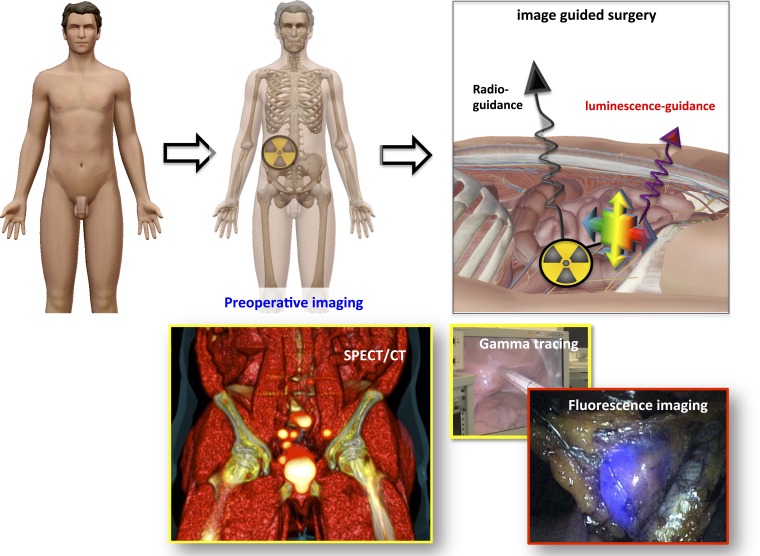
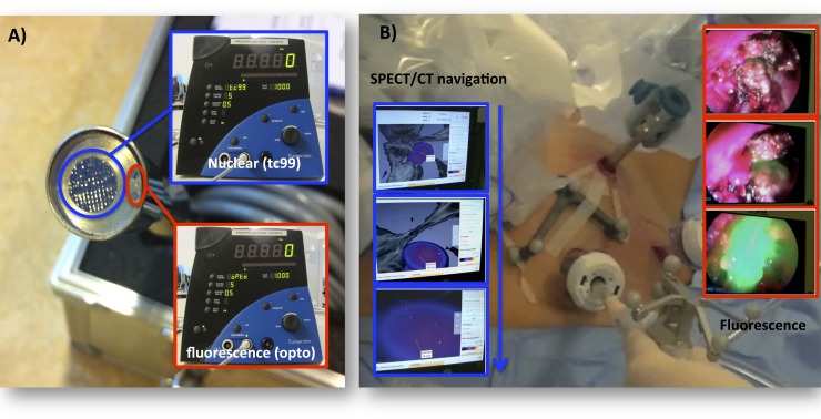
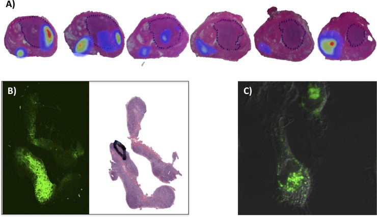
With the evolution of imaging technologies and tracers, the applications for nuclear molecular imaging are growing rapidly. For example, nuclear medicine is increasingly being used to guide surgical resections in complex anatomical locations. Here, a future workflow is envisioned that uses a combination of pre-operative diagnostics, navigation and intraoperative guidance. Radioguidance can provide means for pre-operative and intraoperative identification of "hot" lesions, forming the basis of a virtual data set that can be used for navigation. Luminescence guidance has shown great potential in the intraoperative setting by providing optical feedback, in some cases even in real time. Both of these techniques have distinct drawbacks, which include inaccuracy in areas that contain a background signal (radioactivity) or a limited degree of signal penetration (luminescence). We, and others, have reasoned that hybrid/multimodal approaches that integrate the use of these complementary modalities may help overcome their individual weaknesses. Ultimately, this will lead to advancement of the field of interventional molecular imaging/image-guided surgery. In this review, an overview of clinically applied hybrid surgical guidance technologies is given, whereby the focus is placed on tracers and hardware.


Figures

Figure 1.
Figure 1.
Emissions and their degree of tissue penetration. (a) A schematic representation on the relative ability of radioactive (α-, β- and γ-emissions) and luminescence emissions (Cerenkov luminescence and fluorescence) to penetrate human tissue. (b) Schematics of a phantom setup, where (1) represents a gel-phantom with absorbance properties similar to human tissue and (2) represents a diagonally placed glass rod containing a radioisotope [technetium-99m (99mTc)] and a fluorescence dye (Cy5). The underlying graph depicts the quantified signal intensities along the dotted line as derived from top-view images (data adapted from Oosterom et al published under the terms of the Creative Common Attribution 4.0 unported license http://creativecommons.org/licenses/by/4.0). The anatomical image was made using the visual body software package (Human Anatomy Atlas v. 3.0.1; Argosy Publishing, Inc., Newton, MA).
Figure 2.
Figure 2.
The hybrid surgical guidance concept. The top line displays a schematic setup that illustrates the hybrid surgical guidance concept. The bottom example depicts the use of indocyanine green–technetium-99m–nanocolloid during robot-assisted SN biopsy in a patient with prostate cancer: single-photon emission CT (SPECT)/CT imaging followed by intraoperative gamma tracing and endoscopic fluorescence imaging. The anatomical image was made using the visual body software package (Human Anatomy Atlas v. 3.0.1; Argosy Publishing, Inc., Newton, MA).
Figure 3.
Figure 3.
Illustration of hybrid modalities in use for surgical guidance. (a) An optonuclear technology that allows for both gamma and fluorescence tracing. (b) The surgical navigation of a fluorescence laparoscope in pre-operative single-photon emission CT (SPECT)/CT images.
Figure 4.
Figure 4.
The use of fluorescence in ex vivo/pathological specimens and cells. (a) An example that illustrates how indocyanine green–technetium-99m–nanocolloid (ICG-99mTc-nanocolloid) deposits (in rainbow) are distributed throughout the prostate in relation to the tumour distribution (dotted line: adapted from Buckle et al with permission form the Society of Nuclear Medicine and Molecular Imaging Inc.). (b) Microscopic imaging of ICG-99mTc-nanocolloid uptake in a sentinel node: fluorescence (green) and relative to the tumour distribution (black line: adapted from Brouwer et al with permission from Elsevier). (c) Confocal image of Cy5-nanocolloid (blue) in human macrophages.

Similar articles

Cited by

References

    1. Amersi F, Hansen NM. The benefits and limitations of sentinel lymph node biopsy. Curr Treat Options Oncol 2006; 7: 141–51. doi: 10.1007/s11864-006-0049-y - DOI - PubMed
    1. Nieweg OE, Estourgie SH, van Rijk MC, Kroon BB. Rationale for superficial injection techniques in lymphatic mapping in breast cancer patients. J Surg Oncol 2004; 87: 153–6. doi: 10.1002/jso.20108 - DOI - PubMed
    1. Bluemel C, Herrmann K, Giammarile F, Nieweg OE, Dubreuil J, Testori A, et al. . EANM practice guidelines for lymphoscintigraphy and sentinel lymph node biopsy in melanoma. Eur J Nucl Med Mol Imaging 2015; 42: 1750–66. doi: 10.1007/s00259-015-3135-1 - DOI - PubMed
    1. Chin PT, Welling MM, Meskers SC, Valdes Olmos RA, Tanke H, van Leeuwen FW. Optical imaging as an expansion of nuclear medicine: Cerenkov-based luminescence vs fluorescence-based luminescence. Eur J Nucl Med Mol Imaging 2013; 40: 1283–91. doi: 10.1007/s00259-013-2408-9 - DOI - PubMed
    1. van Leeuwen FW, van der Poel HG. Surgical guidance in prostate cancer: “from molecule to man” translations. Clin Cancer Res 2015. Epub ahead of print . doi: 10.1158/1078-0432.CCR-15-2575 - DOI - PubMed

MeSH terms