Anti-TNF-α treatment modulates SASP and SASP-related microRNAs in endothelial cells and in circulating angiogenic cells
- PMID: 26943583
- PMCID: PMC4914260
- DOI: 10.18632/oncotarget.7858
Anti-TNF-α treatment modulates SASP and SASP-related microRNAs in endothelial cells and in circulating angiogenic cells
Abstract
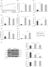
Endothelial cell senescence is characterized by acquisition of senescence-associated secretory phenotype (SASP), able to promote inflammaging and cancer progression. Emerging evidence suggest that preventing SASP development could help to slow the rate of aging and the progression of age-related diseases, including cancer. Aim of this study was to evaluate whether and how adalimumab, a monoclonal antibody directed against tumor necrosis factor-α (TNF-α), a major SASP component, can prevent the SASP. A three-pronged approach has been adopted to assess the if adalimumab is able to: i) modulate a panel of classic and novel senescence- and SASP-associated markers (interleukin [IL]-6, senescence associated-β-galactosidase, p16/Ink4a, plasminogen activator inhibitor 1, endothelial nitric oxide synthase, miR-146a-5p/Irak1 and miR-126-3p/Spred1) in human umbilical vein endothelial cells (HUVECs); ii) reduce the paracrine effects of senescent HUVECs' secretome on MCF-7 breast cancer cells, through wound healing and mammosphere assay; and iii) exert significant decrease of miR-146a-5p and increase of miR-126-3p in circulating angiogenic cells (CACs) from psoriasis patients receiving adalimumab in monotherapy.TNF-α blockade associated with adalimumab induced significant reduction in released IL-6 and significant increase in eNOS and miR-126-3p expression levels in long-term HUVEC cultures.A significant reduction in miR-146a-5p expression levels both in long-term HUVEC cultures and in CACs isolated from psoriasis patients was also evident. Interestingly, conditioned medium from senescent HUVECs treated with adalimumab was less consistent than medium from untreated cells in inducing migration- and mammosphere- promoting effects on MCF-7 cells.Our findings suggest that adalimumab can induce epigenetic modifications in cells undergoing senescence, thus contributing to the attenuation of SASP tumor-promoting effects.
Keywords: Gerotarget; HUVEC; SASP; miR-126-3p; miR-146a-5p; replicative senescence.
Conflict of interest statement
None of the authors have competing interests.
Figures


Similar articles
-
Anti-inflammatory effect of ubiquinol-10 on young and senescent endothelial cells via miR-146a modulation.Free Radic Biol Med. 2013 Oct;63:410-20. doi: 10.1016/j.freeradbiomed.2013.05.033. Epub 2013 May 30. Free Radic Biol Med. 2013. PMID: 23727324
-
MiR-146a as marker of senescence-associated pro-inflammatory status in cells involved in vascular remodelling.Age (Dordr). 2013 Aug;35(4):1157-72. doi: 10.1007/s11357-012-9440-8. Epub 2012 Jun 13. Age (Dordr). 2013. PMID: 22692818 Free PMC article.
-
Senescence-associated microRNAs target cell cycle regulatory genes in normal human lung fibroblasts.Exp Gerontol. 2017 Oct 1;96:110-122. doi: 10.1016/j.exger.2017.06.017. Epub 2017 Jun 27. Exp Gerontol. 2017. PMID: 28658612
-
Mitochondrial (Dys) Function in Inflammaging: Do MitomiRs Influence the Energetic, Oxidative, and Inflammatory Status of Senescent Cells?Mediators Inflamm. 2017;2017:2309034. doi: 10.1155/2017/2309034. Epub 2017 Dec 27. Mediators Inflamm. 2017. PMID: 29445253 Free PMC article. Review.
-
MicroRNA-34a: the bad guy in age-related vascular diseases.Cell Mol Life Sci. 2021 Dec;78(23):7355-7378. doi: 10.1007/s00018-021-03979-4. Epub 2021 Oct 26. Cell Mol Life Sci. 2021. PMID: 34698884 Free PMC article. Review.
Cited by
-
MicroRNAs are critical regulators of senescence and aging in mesenchymal stem cells.Bone. 2021 Jan;142:115679. doi: 10.1016/j.bone.2020.115679. Epub 2020 Oct 3. Bone. 2021. PMID: 33022453 Free PMC article. Review.
-
Cellular senescence and SASP in tumor progression and therapeutic opportunities.Mol Cancer. 2024 Aug 31;23(1):181. doi: 10.1186/s12943-024-02096-7. Mol Cancer. 2024. PMID: 39217404 Free PMC article. Review.
-
Breast Tissue Biology Expands the Possibilities for Prevention of Age-Related Breast Cancers.Front Cell Dev Biol. 2019 Aug 28;7:174. doi: 10.3389/fcell.2019.00174. eCollection 2019. Front Cell Dev Biol. 2019. PMID: 31555644 Free PMC article. Review.
-
Senescent cell clearance by the immune system: Emerging therapeutic opportunities.Semin Immunol. 2018 Dec;40:101275. doi: 10.1016/j.smim.2019.04.003. Epub 2019 May 11. Semin Immunol. 2018. PMID: 31088710 Free PMC article. Review.
-
Age-related immune alterations and cerebrovascular inflammation.Mol Psychiatry. 2022 Feb;27(2):803-818. doi: 10.1038/s41380-021-01361-1. Epub 2021 Oct 28. Mol Psychiatry. 2022. PMID: 34711943 Free PMC article. Review.
References
-
- Franceschi C, Bonafè M, Valensin S, Olivieri F, De Luca M, Ottaviani E, De Benedictis G. Inflamm-aging: an evolutionary perspective on immunosenescence. Ann N Y Acad Sci. 2000;908:244–54. - PubMed
-
- Campisi J, d'Adda di Fagagna F. Cellular senescence: when bad things happen to good cells. Nat Rev Mol Cell Biol. 2007;8:729–40. - PubMed
MeSH terms
Substances
LinkOut - more resources
Full Text Sources
Other Literature Sources
Medical
Molecular Biology Databases
Miscellaneous

