. 2016 Mar 4;11(3):e0150784.
doi: 10.1371/journal.pone.0150784. eCollection 2016.

Characterization and Localization of Citrullinated Proteoglycan Aggrecan in Human Articular Cartilage

Affiliations

Characterization and Localization of Citrullinated Proteoglycan Aggrecan in Human Articular Cartilage

Tibor T Glant et al. PLoS One. .

Abstract

Background: Rheumatoid arthritis (RA) is an autoimmune disease of the synovial joints. The autoimmune character of RA is underscored by prominent production of autoantibodies such as those against IgG (rheumatoid factor), and a broad array of joint tissue-specific and other endogenous citrullinated proteins. Anti-citrullinated protein antibodies (ACPA) can be detected in the sera and synovial fluids of RA patients and ACPA seropositivity is one of the diagnostic criteria of RA. Studies have demonstrated that RA T cells respond to citrullinated peptides (epitopes) of proteoglycan (PG) aggrecan, which is one of the most abundant macromolecules of articular cartilage. However, it is not known if the PG molecule is citrullinated in vivo in human cartilage, and if so, whether citrulline-containing neoepitopes of PG (CitPG) can contribute to autoimmunity in RA.

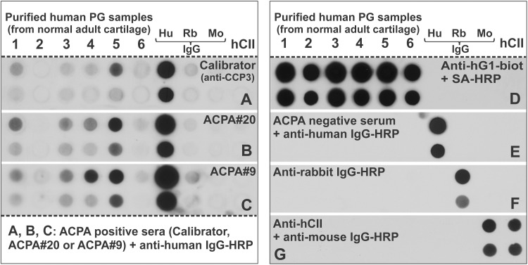
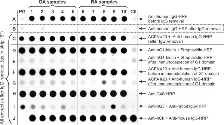
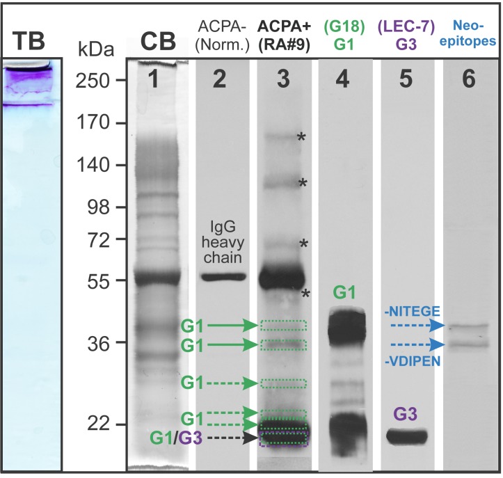
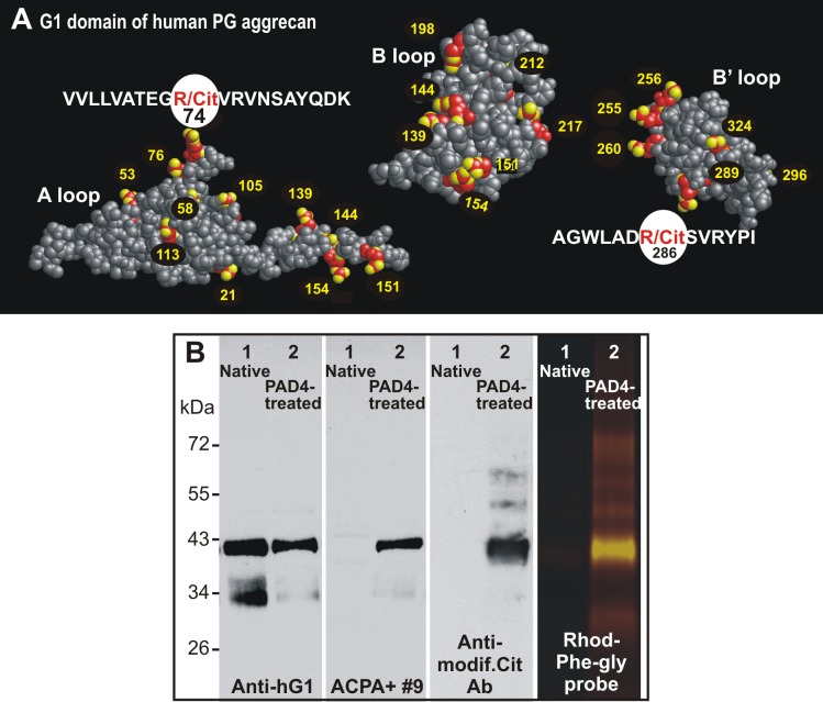
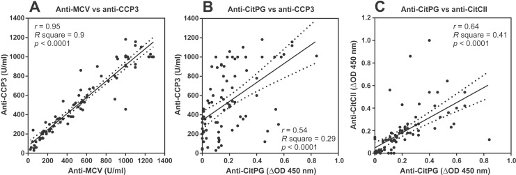
Methods: CitPG was detected in human cartilage extracts using ACPA+ RA sera in dot blot and Western blot. Citrullination status of in vitro citrullinated recombinant G1 domain of human PG (rhG1) was confirmed by antibody-based and chemical methods, and potential sites of citrullination in rhG1 were explored by molecular modeling. CitPG-specific serum autoantibodies were quantified by enzyme-linked immunosorbent assays, and CitPG was localized in osteoarthritic (OA) and RA cartilage using immunohistochemistry.

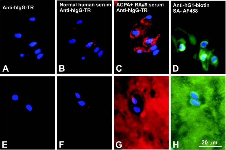
Findings: Sera from ACPA+ RA patients reacted with PG purified from normal human cartilage specimens. PG fragments (mainly those containing the G1 domain) from OA or RA cartilage extracts were recognized by ACPA+ sera but not by serum from ACPA- individuals. ACPA+ sera also reacted with in vitro citrullinated rhG1 and G3 domain-containing fragment(s) of PG. Molecular modeling suggested multiple sites of potential citrullination within the G1 domain. The immunohistochemical localization of CitPG was different in OA and RA cartilage.

Conclusions: CitPG is a new member of citrullinated proteins identified in human joints. CitPG could be found in both normal and diseased cartilage specimens. Antibodies against CitPG may trigger or augment arthritis by forming immune complexes with this autoantigen in the joints of ACPA+ RA patients.


Conflict of interest statement

Competing Interests: The authors have declared that no competing interests exist.

Figures

Fig 1
Fig 1. Recognition of proteoglycan (PG) aggrecan purified from normal human cartilage by ACPA-positive human sera.
(A-C) Dots of human PG aggrecan that was purified from 6 normal cartilage samples (1–6) were applied to nitrocellulose membranes (upper dots: 2 μg, lower dots: 0.2 μg PG) along with various control IgGs purified from human (Hu), rabbit (Rb) or mouse (Mo) serum (upper dots: 20 ng, lower dots: 2 ng IgG) and human type II collagen (hCII) purified from normal cartilage (upper dot: 10 μg, lower dot: 1 μg hCII). The membranes were subjected to immunostaining with ACPA-positive sera including the (A) “Calibrator” serum from the anti-CCP3 assay kit and (B, C) sera from two ACPA+ RA patients (#20 and #9), followed by horseradish peroxidase (HRP)-labeled anti-human IgG. (D-G) Specificity controls included blotting with (D) a biotinylated monoclonal antibody (mAb) specific to human PG G1 domain (anti-hG1-biot) followed by streptavidin (SA)-HRP, (E) ACPA- serum followed by anti-human IgG-HRP, (F) anti-rabbit IgG-HRP, and (G) mouse antibody to hCII (anti-hCII) followed by anti-mouse IgG-HRP.
Fig 2
Fig 2. Recognition of PG in crude extracts of osteoarthritis (OA) and rheumatoid arthritis (RA) cartilage specimens by ACPA and PG-specific antibodies.
Dots of crude extracts of cartilage from OA donors (row A, samples 1–5) and RA donors (samples 6–10) were applied to a nitrocellulose membrane strip. Purified human PG and CII (far left and far right dots, respectively) served as controls. Blotting with the secondary antibody (anti-human IgG-HRP) revealed positive reactions (row A) with all crude extracts, but not with purified hPG or hCII. The “positive” reaction disappeared after the contaminating IgG was removed from the crude extracts (row B). These IgG-free cartilage extracts were used for subsequent dot blots. The ACPA+#20 serum (row A) reacted with all cartilage extracts and purified PG (row C). Similarly, the G1 domain-specific mAb recognized purified PG and PG in the crude extracts (row D), but no reaction was detected when the G1 domain was removed by immune absorption (row E). The reactivity of the cartilage extracts with ACPA+#20 serum (row F) was nearly completely lost after G1 domain immunodepletion (row G). As demonstrated by the anti-chondroitin 4-sulfate (C4S)-specific antibody, PG in all extracts and in the purified PG sample were glycosylated (row H). Some extracts and the purified PG contained small amounts of the PG G3 domain (row I), and all crude extracts contained cartilage-specific CII (row J).
Fig 3
Fig 3. Western blots to identify PG domains and fragments of OA cartilage extract recognized by ACPA-positive serum.
The crude extract of OA cartilage (shown as sample 4 in Fig 2) was loaded onto 8% SGS-PAGE gel and stained with toluidine blue (TB) to visualize PG GAG chains. Essentially all of the TB-positive PG material remained in the stacking gel. To facilitate resolution, chondroitin sulfate chains were removed by digestion with chondroitinase ABC, and aliquots of the deglycosylated extract was loaded onto 6 lanes of a SDS-PAGE gel. Coomassie blue (CB) staining of the gel (lane 1) showed good resolution of the proteins of the OA cartilage extract after deglycosylation. Following transfer onto a nitrocellulose membrane, vertical strips of the membrane were probed with human sera or PG-specific antibodies (lanes 2–6). Immunostaining with ACPA- (normal) serum followed by anti-human IgG-HRP revealed a single protein band most likely corresponding to the heavy chain of contaminating IgG (lane 2). The ACPA+ serum detected several additional bands (lane 3). To identify these bands, replicate strips of the membrane were probed with antibodies against the G1 or G3 domain of PG and a pair of antibodies recognizing protease-generated PG neoepitopes. The respective antibodies showed reactions with the G1 (lane 4), G3 (lane 5) as well as with the neoepitopes -NITEGE and -VDIPEN (lane 6). There were additional bands above 55 kDa (depicted with asterisks in lane 3) that could not be identified as PG fragments. One representative sample of over 10 Western blots (using different crude extracts and ACPA+ sera) is shown.
Fig 4
Fig 4. Immunohistochemical localization of ACPA-reactive (citrullinated) epitopes in OA and RA cartilage sections.
(A-D) Frozen sections of OA knee (tibial plateau) cartilage were immunostained with (A) Texas red-labeled anti-human IgG (anti-hIgG-TR), (B) normal (ACPA-negative) human serum followed by anti-hIgG-TR, (C) ACPA+ serum (RA#9) followed by anti-hIgG-TR, or (D) a biotinylated anti-human G1 antibody followed by Alexa Fluor 488-labeled streptavidin (SA-AF488). (E-H) Frozen sections prepared from the RA tibial plateau cartilage were immunostained with the same sera and antibodies as listed for A-D. Cell nuclei in all sections were visualized by DAPI staining. (A and E) Anti-hIgG-TR alone did not stain the sections, and (B and F) negligible reaction (red fluorescence) was observed when the tissues were first stained with ACPA- serum. (C) The ACPA+ serum primarily stained the chondrocyte pericellular matrix in the OA cartilage, but (G) it diffusely stained the entire matrix of the RA cartilage. Similar staining patters to those with ACPA+ serum were observed when (D) the OA and (H) RA cartilage sections were incubated with biotinylated anti-hG1 mAb (green fluorescence), suggesting at least partial co-localization of PG G1 and citrullinated epitopes in both OA and RA cartilage.
Fig 5
Fig 5. Positions of arginine residues within the three-dimensional structure of the human G1 domain and detection of citrullinated rhG1.
(A) 3-D images of the A, B, and B’ loops of the G1 domain illustrate the location of arginine (R) residues (red-yellow balls with numbers) and the two previously reported immunodominant epitopes (sequences highlighted), both of which contain arginine residues that may become citrulline (R/Cit) [11][12][13]. The three loops of G1 were rotated relative to each other using RasMol software in order to expose the R-rich surfaces. The amino acid sequence is shown in S1 Fig. (B) In vitro citrullination of rhG1 was performed using PAD4 enzyme. The native (lanes 1) and PAD4-treated (lanes 2) rhG1 proteins were loaded onto SDS-PAGE gels and transferred to nitrocellulose membranes. Two of the membranes were probed with either anti-hG1 mAb: (first panel) or with ACPA+ RA serum (second panel). The citrulline residues present in the same proteins on the third membrane were subjected to chemical modification and then probed with an Ab specific for chemically-modified citrulline (anti-modif. Cit Ab, third panel). Native and PAD4-treated rhG1 proteins were also reacted with citrulline-specific phenylglyoxal conjugated with rhodamine (Rhod-Phe-Gly) and subjected to SDS-PAGE (fourth panel). While the anti-hG1 mAb reacted with both the native and citrullinated forms of the protein, only the citrullinated hG1 was detected by ACPA, the anti-modified citrulline Ab, and the phenylglyoxal probe.
Fig 6
Fig 6. Correlations between ACPA of different specificities including citrullinated PG in the sera of RA patients.
(A) Concentrations of antibodies to mutated citrullinated vimentin (MCV) and cyclic citrullinated peptides (CCP) were measured in serum samples from 84 RA patients using commercial ELISA kits. Results were expressed as units/ml (U/ml). Correlation analysis revealed strong positive correlation between anti-MCV and anti-CCP3 levels (r = 0.95, R square = 0.9, p<0.0001). (B) Concentrations of anti-CitPG antibodies were measured by in-house ELISA using in vitro citrullinated rhG1 (shown in Fig 5B) as antigen (CitPG). The results were expressed as delta optical density (ΔOD, the OD values of anti-CitPG minus the OD values of anti-PG as described in the Methods). The ΔOD values were correlated with the concentrations (U/ml) of anti-CCP in the same 84 RA serum samples. (C) Purified hCII was also citrullinated by PAD4, and CitCII was used to measure anti-CitCII levels in the 84 RA serum samples by ELISA. The dotted lines indicate the 95% confidence intervals.

Similar articles

Cited by

References

    1. Fox DA. Etiology and Pathogenesis of Rheumatoid Arthritis In: Koopman WJ, Moreland LW, editors. Arthritis and Allied Conditions: A Textbook of Rheumatology. Philadelphia: Lippincott Williams & Wilkins; 2005. pp. 1085–1102.
    1. Firestein GS. Rheumatoid arthritis: Etiology and pathogeneis of rheumatoid arthritis In: Ruddy S, Harris ED, Sledge CB, Kelley WN, editors. Kelley's Textbook of Rheumatology. Philadelphia, PA: W.B.Saunders Co.; 2005. pp. 996–1045.
    1. Schellekens GA, de Jong BA, van den Hoogen FH, van de Putte LB, van Venrooij WJ. Citrulline is an essential constituent of antigenic determinants recognized by rheumatoid arthritis-specific autoantibodies. J Clin Invest 1998; 101: 273–281 - PMC - PubMed
    1. Bas S, Perneger TV, Seitz M, Tiercy JM, Roux-Lombard P, Guerne PA. Diagnostic tests for rheumatoid arthritis: comparison of anti-cyclic citrullinated peptide antibodies, anti-keratin antibodies and IgM rheumatoid factors. Rheumatology (Oxford) 2002; 41: 809–814 - PubMed
    1. Trouw LA, Huizinga TW, Toes RE. Autoimmunity in rheumatoid arthritis: different antigens—common principles. Ann Rheum Dis 2013; 72 Suppl 2: ii132–ii136 10.1136/annrheumdis-2012-202349 - DOI - PubMed

Publication types

MeSH terms