

Integrally calcified solitary fibrous tumor in the retroperitoneum: a case report and review of the literature
- PMID: 26943690
- PMCID: PMC4752942
- DOI: 10.1186/s40792-016-0143-8
Integrally calcified solitary fibrous tumor in the retroperitoneum: a case report and review of the literature
Abstract
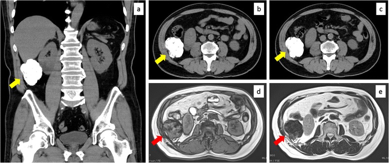
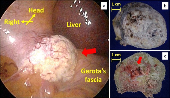
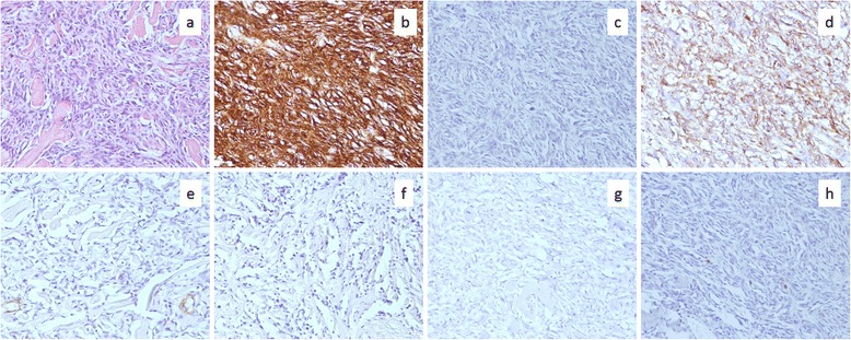
Solitary fibrous tumor (SFT) is a rare stromal neoplasm and usually occurs in the thoracic cavity. We here report a case of retroperitoneal SFT with prominent calcification. A 64-year-old man presented with an incidentally detected retroperitoneal mass in the right upper abdomen. Imaging tests indicated an integrally calcified mass. The lesion was observed for 2 years and laparoscopically resected according to the patient's wish. Microscopically, the mass was mostly occupied by calcification and proliferous spindle cells were scattered with positive CD34 expression. We diagnosed morphologically benign SFT and the patient remained disease-free 1 year after the excision. There has been no report of such integrally calcified SFT. Retroperitoneal SFT is difficult to make a preoperative diagnosis, and careful follow-up after the excision is recommended because morphological malignancy does not always correspond to clinical malignancy.
Keywords: Calcification; Retroperitoneum; Solitary fibrous tumor.
Figures
Similar articles
-
Solitary fibrous tumor in the retroperitoneum: A case report.Int J Surg Case Rep. 2024 Jan;114:109118. doi: 10.1016/j.ijscr.2023.109118. Epub 2023 Dec 11. Int J Surg Case Rep. 2024. PMID: 38159392 Free PMC article.
-
Malignant solitary fibrous tumor in retroperitoneum: A case report and literature review.Medicine (Baltimore). 2017 Mar;96(11):e6373. doi: 10.1097/MD.0000000000006373. Medicine (Baltimore). 2017. PMID: 28296778 Free PMC article.
-
Primary solitary fibrous tumor (SFT) in the retroperitoneum.Urol Oncol. 2008 May-Jun;26(3):254-9. doi: 10.1016/j.urolonc.2007.03.024. Epub 2008 Feb 20. Urol Oncol. 2008. PMID: 18452815
-
Solitary fibrous tumor arising from pelvic retroperitoneum: A report of two cases and a review of the literature.J Obstet Gynaecol Res. 2019 Jul;45(7):1391-1397. doi: 10.1111/jog.13965. Epub 2019 Apr 7. J Obstet Gynaecol Res. 2019. PMID: 30957324 Review.
-
Solitary fibrous tumor of the anterior abdominal wall. A case report and review of the literature.Ann Ital Chir. 2019 Jun 13;8:S2239253X19030068. Ann Ital Chir. 2019. PMID: 31366738 Review.
Cited by
-
Solitary fibrous tumour of gluteus: a case report about an uncommon localization of a rare neoplasm.J Surg Case Rep. 2016 Jun 22;2016(6):rjw111. doi: 10.1093/jscr/rjw111. J Surg Case Rep. 2016. PMID: 27333919 Free PMC article.
-
Solitary fibrous tumors from A to Z: a pictorial review with radiologic-pathologic correlation.Insights Imaging. 2025 May 28;16(1):112. doi: 10.1186/s13244-025-01991-x. Insights Imaging. 2025. PMID: 40437277 Free PMC article.
-
Case report: A rare case of malignant solitary fibrous tumor within the joint cavity with review of the literature.Front Oncol. 2024 Nov 29;14:1463362. doi: 10.3389/fonc.2024.1463362. eCollection 2024. Front Oncol. 2024. PMID: 39678501 Free PMC article.
-
Retroperitoneal fibrous tumor recurring as lung metastases after 10 years: a case report.Surg Case Rep. 2021 May 22;7(1):127. doi: 10.1186/s40792-021-01209-4. Surg Case Rep. 2021. PMID: 34023959 Free PMC article.
-
Mesentery solitary fibrous tumor with postoperative recurrence and sarcomatosis: A case report and review of literature.World J Clin Oncol. 2022 Apr 24;13(4):303-313. doi: 10.5306/wjco.v13.i4.303. World J Clin Oncol. 2022. PMID: 35582654 Free PMC article.
References
-
- Travis William D, Brambilia E, Muller-Hermelink HK, Harris CC. World Health Organization Classification of tumors. Pathology and genetics of tumors of the lung, pleura, thymus and heart. Lyon: IARC Press; 2004. - PubMed
LinkOut - more resources
Full Text Sources
Other Literature Sources
