

Characterization of neuropathology in the HIV-1 transgenic rat at different ages
- PMID: 26943969
- PMCID: PMC4779503
- DOI: 10.1016/j.jneuroim.2016.01.022
Characterization of neuropathology in the HIV-1 transgenic rat at different ages
Abstract
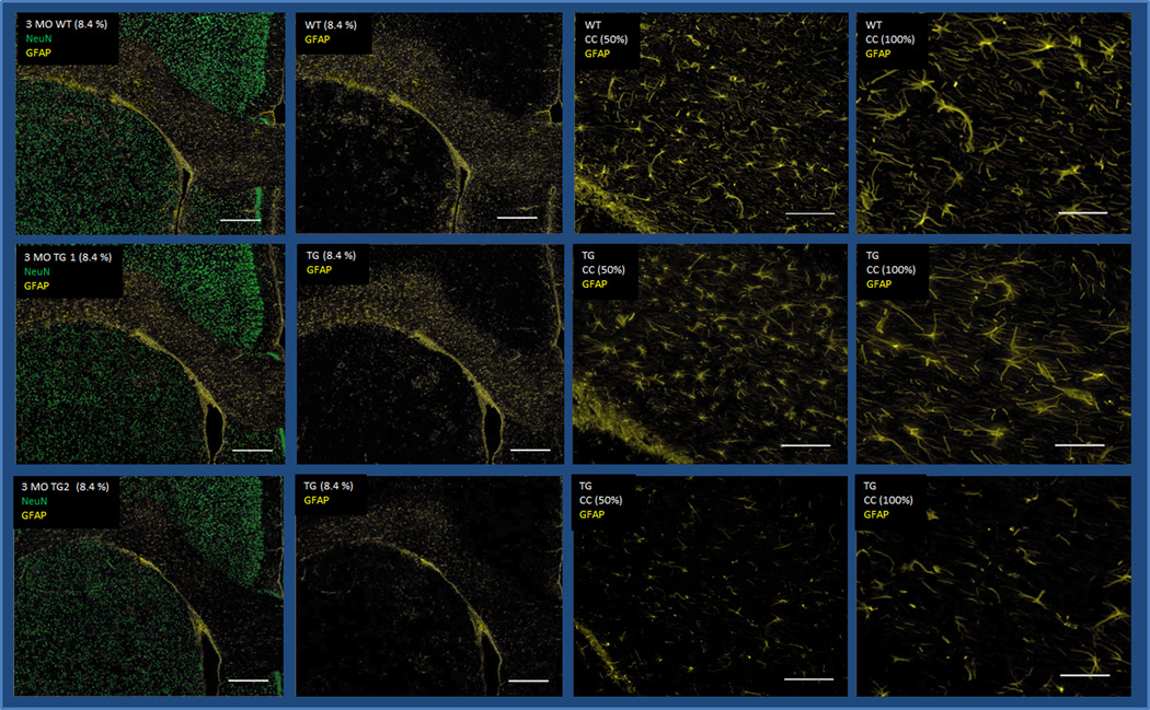
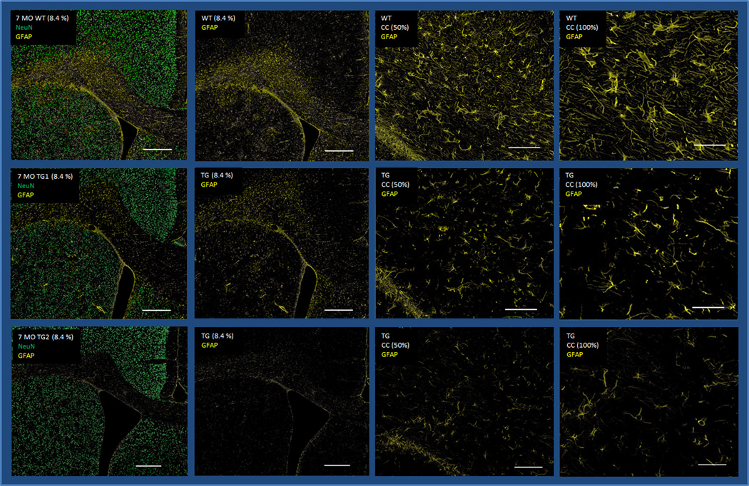
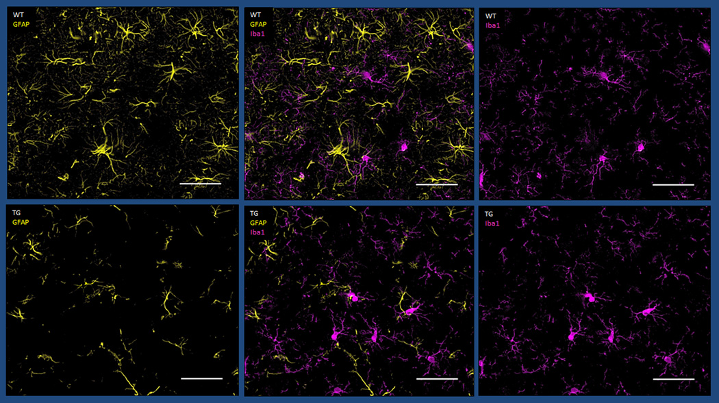
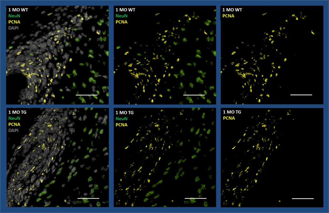
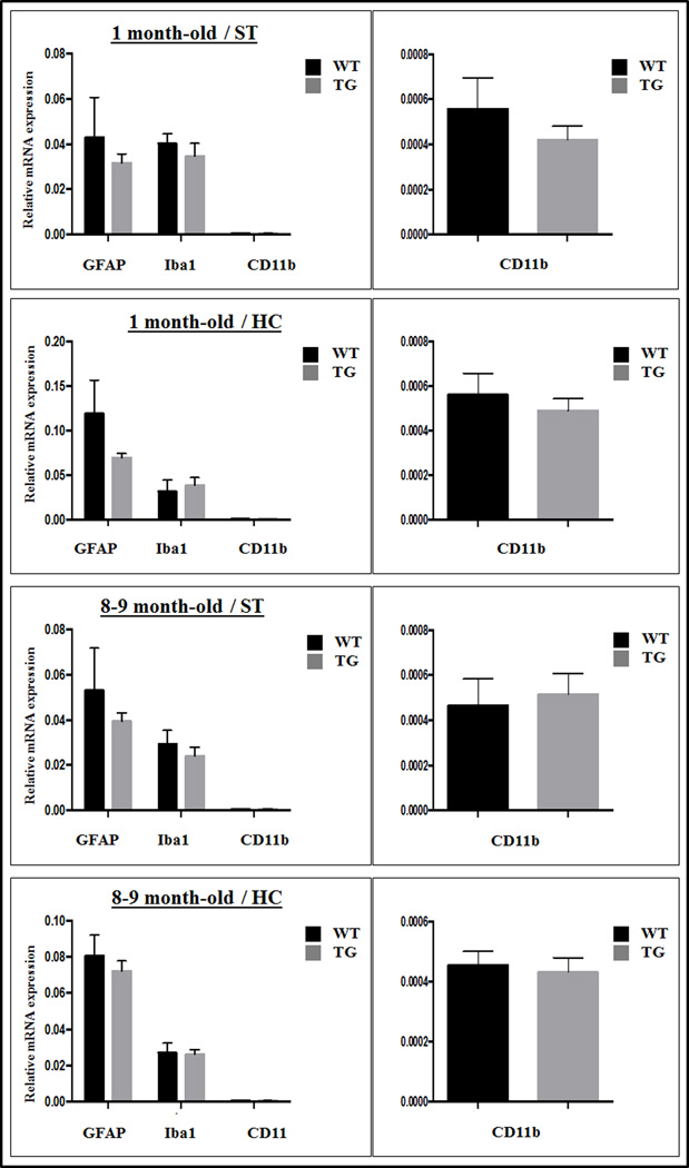
The transgenic HIV-1 rat (Tg) is a commonly used neuroHIV model with documented neurologic/behavioral deficits. Using immunofluorescent staining of the Tg brain, we found astrocytic dysfunction/damage, as well as dopaminergic neuronal loss/dysfunction, both of which worsening significantly in the striatum with age. We saw mild microglial activation in young Tg brains, but this decreased with age. There were no differences in neurogenesis potential suggesting a neurodegenerative rather than a neurodevelopmental process. Gp120 CSF levels exceeded serum gp120 levels in some animals, suggesting local viral protein production in the brain. Further probing of the pathophysiology underlying astrocytic injury in this model is warranted.
Keywords: Astrocytic loss; HIV-1 transgenic rat; Neuro-HIV; Neuropathology; Neurotoxicity; Viral proteins.
Published by Elsevier B.V.
Conflict of interest statement
The authors declare that they have no conflict of interest.
Figures
Similar articles
-
A rat model of human immunodeficiency virus 1 encephalopathy using envelope glycoprotein gp120 expression delivered by SV40 vectors.J Neuropathol Exp Neurol. 2009 May;68(5):456-73. doi: 10.1097/NEN.0b013e3181a10f83. J Neuropathol Exp Neurol. 2009. PMID: 19525894 Free PMC article.
-
Transgenic mice expressing HIV-1 envelope protein gp120 in the brain as an animal model in neuroAIDS research.J Neurovirol. 2018 Apr;24(2):156-167. doi: 10.1007/s13365-017-0584-2. Epub 2017 Oct 26. J Neurovirol. 2018. PMID: 29075998 Free PMC article. Review.
-
Immune activation, viral gene product expression and neurotoxicity in the HIV-1 transgenic rat.J Neuroimmunol. 2012 Jun 15;247(1-2):16-24. doi: 10.1016/j.jneuroim.2012.03.015. Epub 2012 Apr 12. J Neuroimmunol. 2012. PMID: 22503372 Free PMC article.
-
Dysregulation of signal transduction pathways as a potential mechanism of nervous system alterations in HIV-1 gp120 transgenic mice and humans with HIV-1 encephalitis.J Clin Invest. 1996 Feb 1;97(3):789-98. doi: 10.1172/JCI118478. J Clin Invest. 1996. PMID: 8609236 Free PMC article.
-
Transgenic models to assess the neuropathogenic potential of HIV-1 proteins and cytokines.Curr Top Microbiol Immunol. 1995;202:187-205. doi: 10.1007/978-3-642-79657-9_13. Curr Top Microbiol Immunol. 1995. PMID: 7587363 Review. No abstract available.
Cited by
-
Nitrosative Stress Is Associated with Dopaminergic Dysfunction in the HIV-1 Transgenic Rat.Am J Pathol. 2019 Jul;189(7):1375-1385. doi: 10.1016/j.ajpath.2019.03.004. Am J Pathol. 2019. PMID: 31230667 Free PMC article.
-
Brain PET Imaging: Value for Understanding the Pathophysiology of HIV-associated Neurocognitive Disorder (HAND).Curr HIV/AIDS Rep. 2019 Feb;16(1):66-75. doi: 10.1007/s11904-019-00419-8. Curr HIV/AIDS Rep. 2019. PMID: 30778853 Review.
-
Evolution of the HIV-1 transgenic rat: utility in assessing the progression of HIV-1-associated neurocognitive disorders.J Neurovirol. 2018 Apr;24(2):229-245. doi: 10.1007/s13365-017-0544-x. Epub 2017 Jul 20. J Neurovirol. 2018. PMID: 28730408 Free PMC article.
-
Humanized Mice for Infectious and Neurodegenerative disorders.Retrovirology. 2021 Jun 5;18(1):13. doi: 10.1186/s12977-021-00557-1. Retrovirology. 2021. PMID: 34090462 Free PMC article. Review.
-
Lactobacillus rhamnosus GG supernatant enhance neonatal resistance to systemic Escherichia coli K1 infection by accelerating development of intestinal defense.Sci Rep. 2017 Mar 6;7:43305. doi: 10.1038/srep43305. Sci Rep. 2017. PMID: 28262688 Free PMC article.
References
-
- Agrawal L, Louboutin JP, Reyes BA, Van Bockstaele EJ, Strayer DS. HIV-1 tat neurotoxicity: a model of acute and chronic exposure, and neuroprotection by gene delivery of antioxidant enzymes. Neurobiology of Disease. 2012;45:657–670. - PubMed
Publication types
MeSH terms
Substances
Grants and funding
LinkOut - more resources
Full Text Sources
Other Literature Sources
Medical
Miscellaneous
