. 2016 May;15(5):1710-27.
doi: 10.1074/mcp.M116.058131. Epub 2016 Mar 4.

Novel Entropically Driven Conformation-specific Interactions with Tomm34 Protein Modulate Hsp70 Protein Folding and ATPase Activities

Affiliations

Novel Entropically Driven Conformation-specific Interactions with Tomm34 Protein Modulate Hsp70 Protein Folding and ATPase Activities

Michal Durech et al. Mol Cell Proteomics. 2016 May.

Abstract

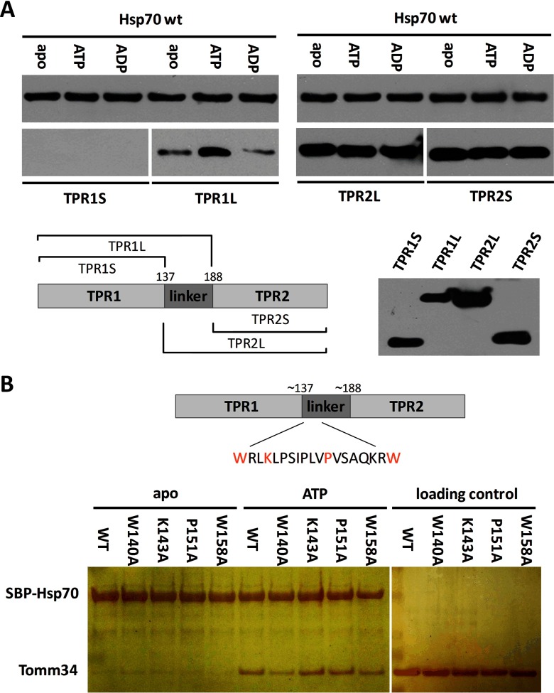
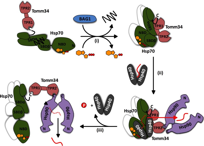
Co-chaperones containing tetratricopeptide repeat (TPR) domains enable cooperation between Hsp70 and Hsp90 to maintain cellular proteostasis. Although the details of the molecular interactions between some TPR domains and heat shock proteins are known, we describe a novel mechanism by which Tomm34 interacts with and coordinates Hsp70 activities. In contrast to the previously defined Hsp70/Hsp90-organizing protein (Hop), Tomm34 interaction is dependent on the Hsp70 chaperone cycle. Tomm34 binds Hsp70 in a complex process; anchorage of the Hsp70 C terminus by the TPR1 domain is accompanied by additional contacts formed exclusively in the ATP-bound state of Hsp70 resulting in a high affinity entropically driven interaction. Tomm34 induces structural changes in determinants within the Hsp70-lid subdomain and modulates Hsp70/Hsp40-mediated refolding and Hsp40-stimulated Hsp70 ATPase activity. Because Tomm34 recruits Hsp90 through its TPR2 domain, we propose a model in which Tomm34 enables Hsp70/Hsp90 scaffolding and influences the Hsp70 chaperone cycle, providing an additional role for co-chaperones that contain multiple TPR domains in regulating protein homeostasis.


Figures

Fig. 1.
Fig. 1.
Tomm34 binding to Hsp70 is ATP-dependent and requires cooperation of both SBD and NBD. Bacterially purified SBP-Hsp70 constructs were immobilized on streptavidin-agarose beads and incubated with gel-filtered HEK293 cell lysates conditioned by the indicated compounds (2 mm MgCl2, 1 mm ATP) (A) or with purified Tomm34 protein in buffer containing 0.2 mm ATP/ADP or no nucleotide (B and C). After washing, bound proteins were eluted by biotin and analyzed by Western blotting. ΔC, deletion of PTIEEVD; V438F, mutant defective in substrate binding; L, total cell lysate.
Fig. 2.
Fig. 2.
Different thermodynamics of Tomm34 binding to Hsp70·ATP and Hsp90α. Tomm34 was injected into a cell containing Hsp70·ATP (A) or Hsp90α (B) for isothermal titration calorimetry. Both chaperones bound to Tomm34 with stoichiometries of ∼1:2 (Tomm34:Hsp70/Hsp90α) and dissociation constants of 0.2 μm (at 15 °C) and 0.96 μm (at 25 °C) for Hsp70·ATP and Hsp90α, respectively. See Table I for the full results.
Fig. 3.
Fig. 3.
Tomm34 binding to Hsp70·ATP is dependent on ATP hydrolysis rate modulated by Hsp40 and Bag-1 co-chaperones and influences chaperone activity. A, SBP-Hsp70·ATP/Tomm34 complexes preformed in the presence of ATP (0.2 mm) were washed in nucleotide-free buffer and incubated at 4 °C for the indicated times before elution by biotin. The level of eluted proteins was analyzed by SDS-PAGE and silver staining. B, equimolar amounts (25 pmol) of SBP-Hsp70, Tomm34, and Hsp40 (as indicated) were mixed in increasing concentrations of ATP, eluted by biotin, and analyzed by Western blotting. C, equimolar amounts (25 pmol) of SBP-Hsp70 and Tomm34 were mixed with increasing amounts of Bag-1 in the presence of ATP (0.2 mm). Eluted proteins were analyzed by Western blotting. D, firefly luciferase was chemically denatured, mixed with Hsp70 (1 μm), Hsp40 (2 μm), ATP (1 mm), and varying Tomm34 concentrations, and recovered luminescence was measured. Negative controls described under “Experimental Procedures” did not exhibit luciferase activity and are not shown. E, ATPase activity of Hsp70 (2 μm) was tested at various Hsp40 and Tomm34 concentrations in malachite green assay. Refolding and ATPase experiments were performed in independent triplicates. Error bars represent S.E.
Fig. 4.
Fig. 4.
ATP-induced allosteric domain docking of Hsp70. A, deuteration level differences of Hsp70 peptides in ATP-bound and nucleotide-free state after 1 h of incubation in deuterated buffer. Numbers at left indicate the Hsp70 peptide fragments; schematic representation at left shows Hsp70 domain constitution; L, interdomain linker; T, C-terminal tail. B, original mass spectra of representative peptide fragments of Hsp70 incubated in non-deuterated buffer (ND) or for 1 h in deuterated buffer in the presence/absence of ATP. C and D, crystal structures of Hsp70 bacterial homologue DnaK in ATP-bound state (C, PDB code 4jne) and in nucleotide-free/ADP state (D, PDB code 2kho). NBD, interdomain linker, SBDβ, and SBDα are shown in yellow, black, marine, and green, respectively. Encircled region (C) shows tight association of IA subdomain of NBD, linker, and SBD loops L2,3/L6,7 (peptides indicated with a star in A) when SBDβ is docked onto NBD (compare with I164D mutant, Fig. 5A). Residues I160 and D526 (human numbering I164 and D529) are highlighted in red in ATP/ADP structures, respectively. The images were created in PyMOL.
Fig. 5.
Fig. 5.
Hsp70 I164D lacks the ability to induce NBD-SBDβ docking upon ATP binding, whereas D529A mutant exhibits destabilized SBDβ subdomain/linker conformation in nucleotide-free state. In contrast to Hsp70/Hop binding, the efficient Hsp70/Tomm34 interaction is dependent on NBD-SBDβ docking. A, deuteration level differences of Hsp70 WT and I164D (or D529A) peptides in ATP-bound (red) or nucleotide-free state (blue) after 1 h of incubation in deuterated buffer. Peptides indicated with a star (left panel) highlight the impaired NBD-SBDβ domain docking of I164D mutant (compare with WT, Fig. 4C). B and C, purified SBP-tagged WT or mutant Hsp70s immobilized on streptavidin-agarose beads were incubated with Tomm34 (B) or Hop (C) in buffer containing 0.2 mm ATP, ADP, or no nucleotide. After washing, eluted proteins were analyzed by Western blotting. ΔC, deletion of PTIEEVD.
Fig. 6.
Fig. 6.
Although Hsp70·ATP protects Tomm34 peptides covering TPR1 domain, Tomm34 binding to Hsp70·ATP induces structural changes at the αA/αB helices in Hsp70 SBD. A, difference in deuteration of Tomm34 in the complex with Hsp70 and Tomm34 alone in the presence (red) or absence (blue) of ATP after 10 min (left) or 3 h (right) of incubation in deuterated buffer. B, deuteration kinetics of selected Tomm34 peptides protected by Hsp70 in the presence of ATP, but not in nucleotide-free state. C, difference in deuteration of Hsp70 in the complex with Tomm34 and Hsp70 alone in ATP-bound (red) or nucleotide-free state (blue) after 10 min or 3 h of incubation in deuterated buffer. Hsp70 peptides 519–529 and 533–543 exhibited consistent protection by Tomm34 in the presence of ATP. D, deuteration kinetics of 519–529/533–543 peptides of Hsp70·ATP protected from deuteration by the presence of Tomm34. E, peptides are indicated in red in the tertiary structure representation of DnaK·ATP (PDB code 4jne) with human Hsp70 numbering. Hsp70 domain constitution is colored similarly as in Fig. 4.
Fig. 7.
Fig. 7.
Peptides covering TPR1 and TPR2B domain of Hop are consistently protected by Hsp70. A, difference in deuteration of Hop in the complex with Hsp70 and Hop alone after 30 min of incubation in deuterated buffer. B, deuteration kinetics of selected Hop peptides shows stable protection by Hsp70. C, effect of Hop binding on deuteration of Hsp70 peptides after 30 min of incubation in deuterated buffer.
Fig. 8.
Fig. 8.
Deletion of 533–543 region severely decreased EEVD/ATP-dependent Hsp70/Tomm34 interaction without abrogating allosteric domain docking. However, the deletion has led to destabilization of the α-helical bundle. A, crystal structure of human SBD (PDB code 4po2). SBDβ, SBDα, and 533–543 regions are shown in marine, green, and red, respectively. The lid-SBDβ positioning ionic contacts (D529–R447 and E543–R469) and residues important for structural integrity of α-helical bundle by forming ionic contacts (E534–K589) and hydrophobic core (L542 shown in red, V577, W580, L581, F592, K595, and L599 shown as blue spheres) are indicated. B, deuteration level differences of Hsp70 WT and Δ533–543 peptides in ATP-bound (red) or nucleotide-free state (blue) after 1 h of incubation in deuterated buffer. C and D, purified SBP-Hsp70 WT or Δ533–543 immobilized on streptavidin-agarose beads were incubated with Tomm34 (C) or Hop (D) in buffer containing 0.2 mm ATP, ADP, or no nucleotide. Eluted proteins were analyzed by Western blotting. ΔC, deletion of PTIEEVD.
Fig. 9.
Fig. 9.
Interdomain linker of Tomm34 represents important region for conformation-specific Hsp70/Tomm34 interaction. The length of TPR domain constructs and mutated residues in the linker region are indicated. Purified SBP-Hsp70 WT was preincubated with streptavidin-agarose beads and mixed with TPR constructs (A) or Tomm34 mutants (B) in different nucleotide conditions. After washing, eluted proteins were subjected to SDS-PAGE and Western blot with mouse polyclonal sera against Tomm34 (A) or silver staining (B). The capacity of polyclonal sera to detect purified TPR domains was tested in A.
Fig. 10.
Fig. 10.
Proposed model of Tomm34 role in chaperone-mediated preprotein transport. ADP-bound Hsp70 with entrapped substrate interacts with Tomm34 weakly through EEVD motif. Bag-1 (NEF) accelerates ADP/ATP exchange and thus enhances formation of Hsp70·ATP/Tomm34 complexes (i). Tomm34 binding to Hsp70·ATP interferes with the ability of Hsp40 (32, 84) to deliver preproteins to Hsp70 (ii). Instead, the preproteins are directly transferred to Hsp90 in “open conformation” recruited to the Hsp40/preprotein/Hsp70·ATP/Tomm34 complex by Tomm34 TPR2 (13). Because Hsp90 is thought to recognize near-native conformation of its substrates (88, 89), the delivered preprotein is not effectively processed by Hsp90 chaperone cycle and is released (iii) (90). The released preprotein re-enters the cycle in a semifolded state until its delivery to the destined compartment. The consecutive association of the preprotein with Hsp40, Hsp70, and Hsp90 chaperones prevents its aggregation. The possible Hsp70 dimers are indicated by the shadow lines.

References

    1. Young J. C., Agashe V. R., Siegers K., and Hartl F. U. (2004) Pathways of chaperone-mediated protein folding in the cytosol. Nat. Rev. Mol. Cell Biol. 5, 781–791 - PubMed
    1. Mayer M. P. (2010) Gymnastics of molecular chaperones. Mol. Cell 39, 321–331 - PubMed
    1. Alvira S., Cuéllar J., Röhl A., Yamamoto S., Itoh H., Alfonso C., Rivas G., Buchner J., and Valpuesta J. M. (2014) Structural characterization of the substrate transfer mechanism in Hsp70/Hsp90 folding machinery mediated by Hop. Nat. Commun. 5, 5484. - PubMed
    1. Kirschke E., Goswami D., Southworth D., Griffin P. R., and Agard D. A. (2014) Glucocorticoid receptor function regulated by coordinated action of the Hsp90 and Hsp70 chaperone cycles. Cell 157, 1685–1697 - PMC - PubMed
    1. Zhang H., Amick J., Chakravarti R., Santarriaga S., Schlanger S., McGlone C., Dare M., Nix J. C., Scaglione K. M., Stuehr D. J., Misra S., and Page R. C. (2015) A bipartite interaction between Hsp70 and CHIP regulates ubiquitination of chaperoned client proteins. Structure 23, 472–482 - PMC - PubMed

MeSH terms

Substances

LinkOut - more resources