. 2016 Apr 29;291(18):9383-94.
doi: 10.1074/jbc.M116.721035. Epub 2016 Mar 4.

Structural Insights into the Transport Mechanism of the Human Sodium-dependent Lysophosphatidylcholine Transporter MFSD2A

Affiliations

Structural Insights into the Transport Mechanism of the Human Sodium-dependent Lysophosphatidylcholine Transporter MFSD2A

Debra Q Y Quek et al. J Biol Chem. .

Abstract

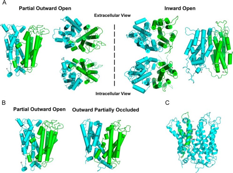
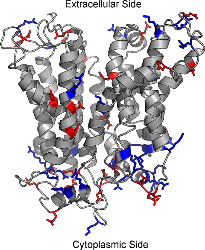
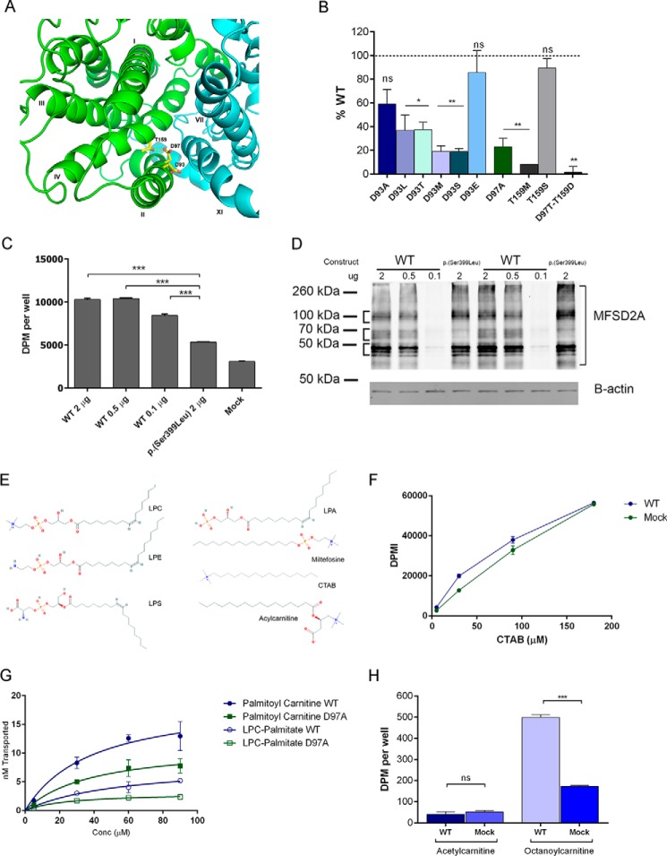
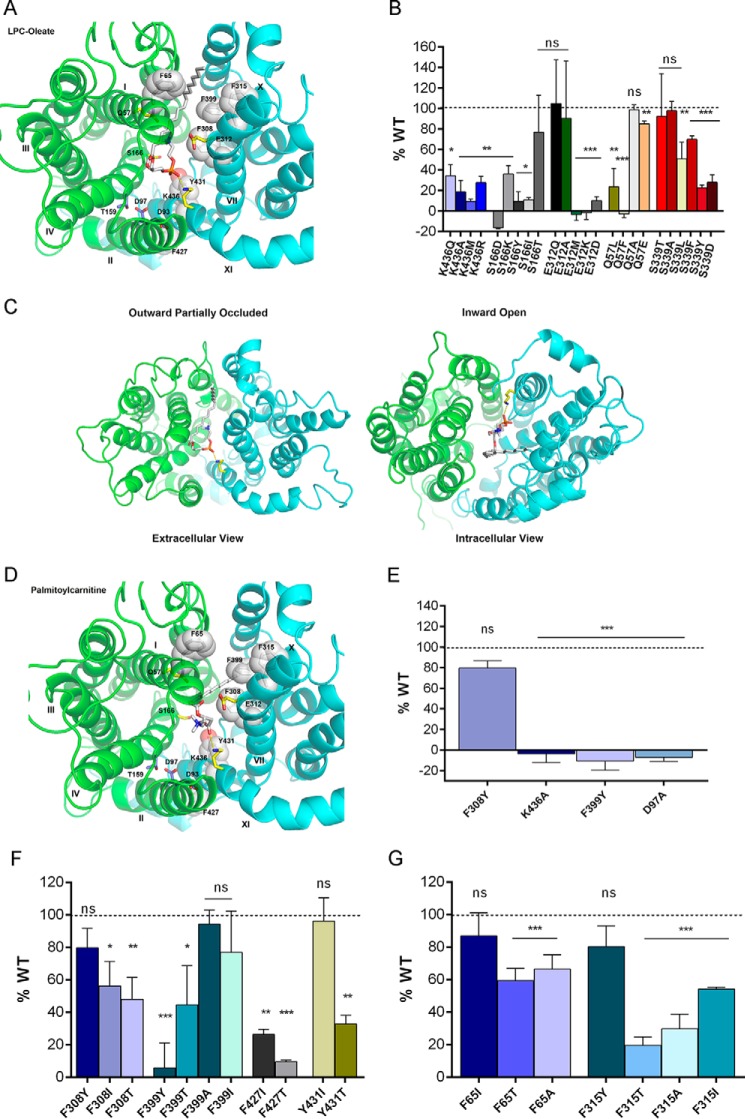
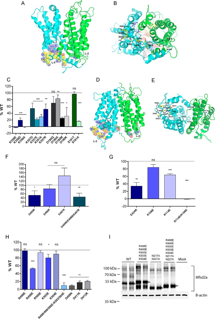
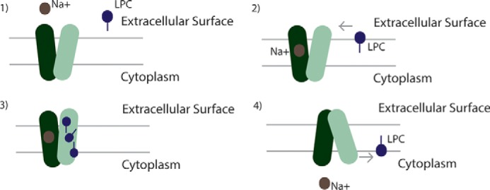
Major facilitator superfamily domain containing 2A (MFSD2A) was recently characterized as a sodium-dependent lysophosphatidylcholine transporter expressed at the blood-brain barrier endothelium. It is the primary route for importation of docosohexaenoic acid and other long-chain fatty acids into fetal and adult brain and is essential for mouse and human brain growth and function. Remarkably, MFSD2A is the first identified major facilitator superfamily member that uniquely transports lipids, implying that MFSD2A harbors unique structural features and transport mechanism. Here, we present three three-dimensional structural models of human MFSD2A derived by homology modeling using MelB- and LacY-based crystal structures and refined by biochemical analysis. All models revealed 12 transmembrane helices and connecting loops and represented the partially outward-open, outward-partially occluded, and inward-open states of the transport cycle. In addition to a conserved sodium-binding site, three unique structural features were identified as follows: a phosphate headgroup binding site, a hydrophobic cleft to accommodate a hydrophobic hydrocarbon tail, and three sets of ionic locks that stabilize the outward-open conformation. Ligand docking studies and biochemical assays identified Lys-436 as a key residue for transport. It is seen forming a salt bridge with the negative charge on the phosphate headgroup. Importantly, MFSD2A transported structurally related acylcarnitines but not a lysolipid without a negative charge, demonstrating the necessity of a negatively charged headgroup interaction with Lys-436 for transport. These findings support a novel transport mechanism by which lysophosphatidylcholines are "flipped" within the transporter cavity by pivoting about Lys-436 leading to net transport from the outer to the inner leaflet of the plasma membrane.

Keywords: X-ray crystallography; blood-brain barrier; brain metabolism; docosahexaenoic acid; drug transport; lysophospholipid; membrane protein; membrane transporter; microcephaly; polyunsaturated fatty acid (PUFA).


Figures

FIGURE 1.
FIGURE 1.
Homology models of MFSD2A. The N- and C-terminal domains are shown in green and cyan, respectively. The helices are labeled with roman numerals. A, overall structure of MFSD2A in the partial outward-open (left of dashed line) and inward-open states (right of dashed line). B, overall structure of MFSD2A in the partial outward-open and outward-partially occluded states. C, rotation of helix VIII in the outward-partially occluded model of MFSD2A to achieve a more likely alignment of the polar side chains of Ser-339, Thr-341, and Thr-343 (labeled in the refined model) toward the protein interior and away from the hydrophobic core of the lipid bilayer. We manually rotated helix VIII in the outward-partially occluded model of MFSD2A. The refined model is shown in cyan and the initial model is shown in green.
FIGURE 2.
FIGURE 2.
Distribution of positive and negative charges in the outward-partially occluded model of MFSD2A. Majority of the charged residues are located at the top and bottom surfaces of the protein, where there is minimal contact with the hydrophobic lipid bilayer, and maximal contact with the hydrophilic extracellular or intracellular environment. There are more positively charged residues on the cytoplasmic surface of MFSD2A. Blue, positively charged residues; red, negatively charged residues.
FIGURE 3.
FIGURE 3.
Sodium-binding site of MFSD2A and the structural requirements for transport. A, sodium-binding site of MFSD2A viewed from the extracellular side of the partial outward-open model with the N- and C-terminal halves in green and cyan, respectively. B, transport of LPC-[14C]oleate after 30 min by HEK293 cells transiently expressing MFSD2A constructs with indicated mutations in residues in the sodium-binding site. Data are expressed as percent activity relative to wild-type MFSD2A. C, transport of LPC-[14C]oleate after 30 min following transfection of HEK293 cells with indicated quantities of WT MFSD2A or S339L plasmids. Mock and S339L transfected cells served as background control and negative control, respectively. D, MFSD2A expression levels in response to concentration of DNA plasmid transfected into HEK293 cells. HEK293 cells were transfected with indicated quantities of plasmid. 18 h after transfection, cells were harvested in RIPA buffer containing protease inhibitor. Western blot was conducted, probing for expression of MFSD2A, and of B-actin as a loading control. S339L was used as a positive control. E, structures of lysophosphatidylcholine (LPC), lysophosphatidylethanolamine (LPE), lysophosphatidylserine (LPS), lysophosphatidic acid (LPA), miltefosine, cetyltrimethylammonium bromide (CTAB), and acylcarnitine. F, concentration-dependent transport of [14C]CTAB after 30 min by wild-type MFSD2A. Mock transfected cells served as a negative control. G, comparison of transport capacity of MFSD2A for LPC-[3H]palmitate and [3H]palmitoylcarnitine. H, transport of 100 μm [3H]acetylcarnitine and [14C]octanoylcarnitine after 20 min by wild-type MFSD2A and mock expressing HEK293 cells. Experiments in B, C, D, and F–H were performed twice in triplicate. Data in B, C, and F–H are expressed as mean ± S.D. *, p < 0.05; **, p < 0.01; ***, p < 0.001; ns, not significant.
FIGURE 4.
FIGURE 4.
Headgroup binding site and hydrophobic cleft of MFSD2A. A, headgroup binding site (yellow sticks) and hydrophobic cleft (white sticks and spheres) viewed from the extracellular side of the outward-partially occluded model. LPC-oleate is shown docked. B, transport of LPC-[14C]oleate after 30 min by HEK293 cells transiently expressing MFSD2A constructs with indicated mutations in residues proposed to be involved in headgroup binding. C, docking of LPC-oleate in the outward-partially occluded and inward-open models. In both models, the negatively charged phosphate group remains in close proximity to the positively charged side chain of Lys-436. In the outward-partially occluded model, the side chain of Lys-436 is pointing outward, and LPC-oleate lies within the translocation pathway with its fatty acyl chain projecting toward the extracellular surface of MFSD2A. In the inward-open model, the side chain of Lys-436 is pointing downwards, whereas LPC-oleate lies within the translocation pathway with its fatty acyl chain projecting toward the cytoplasmic surface of MFSD2A. D, palmitoylcarnitine shown docked as in A, indicating similar residue interactions as seen with LPC-oleate docking. E, transport of [3H]palmitoylcarnitine after 20 min by HEK293 cells transiently expressing MFSD2A constructs with indicated mutations. F and G, transport of LPC-[14C]oleate after 30 min by HEK293 cells transiently expressing MFSD2A constructs with indicated mutations in residues in the proposed hydrophobic cleft. Experiments in B and E–G were performed twice in triplicate. Data in B and E–G are expressed as the mean ± S.D. *, p < 0.05; **, p < 0.01; ***, p < 0.001; ns, not significant.
FIGURE 5.
FIGURE 5.
Ionic locks. A, N- and C-terminal domains are shown in green and cyan, respectively. Three sets of ionic locks are represented as spheres, with lock 1 (L-1) in purple, lock 2 (L-2) in green, and lock 3 (L-3) in yellow in the outward-partially occluded model. B, three sets of ionic locks are shown as sticks, as viewed from the cytoplasmic surface of the outward-partially occluded model, with L-1 in purple, L-2 in green, and L-3 in yellow. C, transport of LPC-[14C]oleate after 30 min by HEK293 cells transiently expressing MFSD2A constructs with indicated mutations in L-1 residues. D, N- and C-terminal domains are shown in green and cyan, respectively. Three sets of ionic locks are represented as spheres, with lock 1 (L-1) in purple, lock 2 (L-2) in green, and lock 3 (L-3) in yellow in the inward-open model. E, three sets of ionic locks are shown as sticks, as viewed from the cytoplasmic surface of the inward-open model, with L-1 in purple, L-2 in green, and L-3 in yellow. In the inward-open model, the residues involved in locks L-1, L-2, and L-3 are not in close proximity as they were in the outward-partially occluded model. F, mutational analysis of residues of L-1. Transport of LPC-[14C]oleate after 30 min by HEK293 cells transiently transfected with MFSD2A constructs with indicated mutations in residues involved in L-1. G, transport of LPC-[14C]oleate after 30 min by HEK293 cells transiently expressing MFSD2A constructs with indicated mutations in L-2 residues. H, transport of LPC-[14C]oleate after 30 min by HEK293 cells transiently expressing MFSD2A constructs with indicated mutations in L-3 residues. I, Western blot analysis of MFSD2A mutants and wild-type MFSD2A indicating a mobility shift in the R498E/R499E/R500E/K503E/K504E mutant relative to wild-type MFSD2A. R498E/R499E/R500E/K503E/K504E mutant with additionally mutated glycosylation sites, N217A/N227A, exhibited a mobility shift relative to non-glycosylated MFSD2A. Arrows indicate MFSD2A bands that have shifted. Experiments in C and F–I were performed twice in triplicate. Data in C and F–H are expressed as the mean ± S.D. *, p < 0.05; **, p < 0.01; ***, p < 0.001; ns, not significant.
FIGURE 6.
FIGURE 6.
Scheme for Na+/LPC symport by MFSD2A. The following four steps in the transport reaction cycle are shown: 1) MFSD2A is in the outward-open conformation; 2) sodium ion(s) bind to the sodium-binding site. LPC inserts into the outer leaflet of the membrane bilayer and diffuses laterally into the central cavity of MFSD2A via a hydrophobic cleft; 3) MFSD2A undergoes conformational changes that pivots the LPC headgroup bound by Lys-436 through the translocation pathway from the outer leaflet to the inner leaflet of the membrane; 4) LPC, being a hydrophobic lipid, exits the transporter laterally into the inner leaflet of the membrane.

References

    1. Salem N. Jr., Litman B., Kim H. Y., and Gawrisch K. (2001) Mechanisms of action of docosahexaenoic acid in the nervous system. Lipids 36, 945–959 - PubMed
    1. Bazan N. G. (2009) Neuroprotectin D1-mediated anti-inflammatory and survival signaling in stroke, retinal degenerations, and Alzheimer's disease. J. Lipid Res. 50, S400–S405 - PMC - PubMed
    1. Baisted D. J., Robinson B. S., and Vance D. E. (1988) Albumin stimulates the release of lysophosphatidylcholine from cultured rat hepatocytes. Biochem. J. 253, 693–701 - PMC - PubMed
    1. Edmond J., Higa T. A., Korsak R. A., Bergner E. A., and Lee W. N. (1998) Fatty acid transport and utilization for the developing brain. J. Neurochem. 70, 1227–1234 - PubMed
    1. Lagarde M., Bernoud N., Brossard N., Lemaitre-Delaunay D., Thiès F., Croset M., and Lecerf J. (2001) Lysophosphatidylcholine as a preferred carrier form of docosahexaenoic acid to the brain. J. Mol. Neurosci. 16, 201–204 - PubMed

Publication types