. 2016 May 15;310(10):H1360-70.
doi: 10.1152/ajpheart.00858.2015. Epub 2016 Mar 4.

Exercise-induced protection against reperfusion arrhythmia involves stabilization of mitochondrial energetics

Affiliations

Exercise-induced protection against reperfusion arrhythmia involves stabilization of mitochondrial energetics

Rick J Alleman et al. Am J Physiol Heart Circ Physiol. .

Abstract

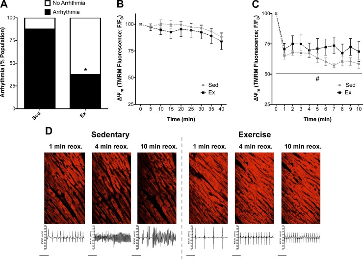
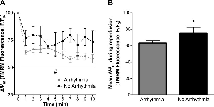
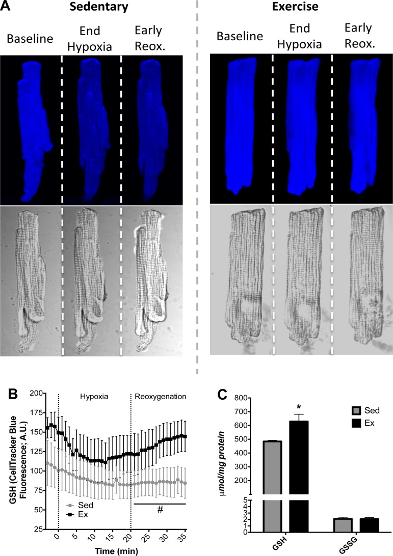
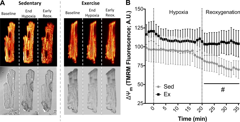
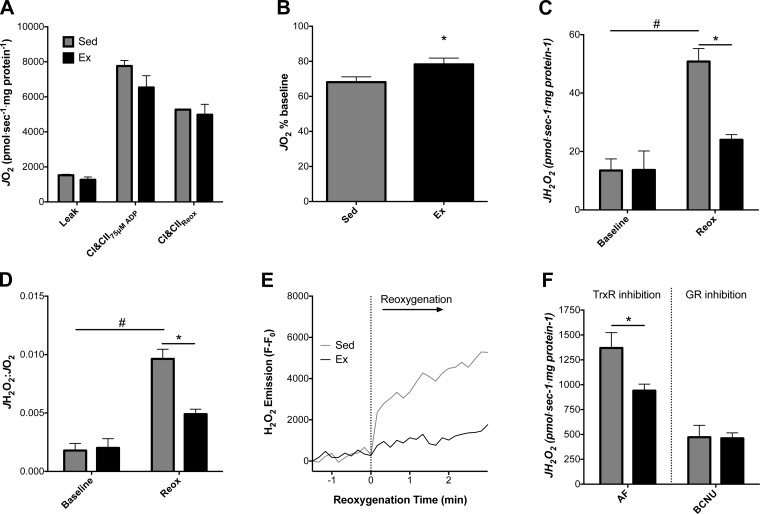
Mitochondria influence cardiac electrophysiology through energy- and redox-sensitive ion channels in the sarcolemma, with the collapse of energetics believed to be centrally involved in arrhythmogenesis. This study was conducted to determine if preservation of mitochondrial membrane potential (ΔΨm) contributes to the antiarrhythmic effect of exercise. We utilized perfused hearts, isolated myocytes, and isolated mitochondria exposed to metabolic challenge to determine the effects of exercise on cardiac mitochondria. Hearts from sedentary (Sed) and exercised (Ex; 10 days of treadmill running) Sprague-Dawley rats were perfused on a two-photon microscope stage for simultaneous measurement of ΔΨm and ECG. After ischemia-reperfusion, the collapse of ΔΨm was commensurate with the onset of arrhythmia. Exercise preserved ΔΨm and decreased the incidence of fibrillation/tachycardia (P < 0.05). Our findings in intact hearts were corroborated in isolated myocytes exposed to in vitro hypoxia-reoxygenation, with Ex rats demonstrating enhanced redox control and sustained ΔΨm during reoxygenation. Finally, we induced anoxia-reoxygenation in isolated mitochondria using high-resolution respirometry with simultaneous measurement of respiration and H2O2 Mitochondria from Ex rats sustained respiration with lower rates of H2O2 emission than Sed rats. Exercise helps sustain postischemic mitochondrial bioenergetics and redox homeostasis, which is associated with preserved ΔΨm and protection against reperfusion arrhythmia. The reduction of fatal ventricular arrhythmias through exercise-induced mitochondrial adaptations indicates that mitochondrial therapeutics may be an effective target for the treatment of heart disease.

Keywords: arrhythmia; cardioprotection; exercise; membrane potential; mitochondria.


Figures

Fig. 1.
Fig. 1.
Arrhythmia and simultaneous 2-photon imaging of mitochondrial membrane potential (ΔΨm) in isolated hearts during ischemia-reperfusion. A: percentage of hearts from exercised (Ex) and sedentary (Sed) rats that transitioned to arrhythmia (ventricular tachycardia/ventricular fibrillation) following 40 min of ischemia. B and C: baseline tetramethylrhodamine methyl ester (TMRM) fluorescence (ΔΨm) values were used to normalize all data (F/F0) during ischemia (B) and reperfusion (C). Values (means ± SE) are expressed as percentage of the population for arrhythmia (n = 7–8 per group). *P < 0.05 vs. Sed; #P < 0.05 vs. Sed main effect. D: representative images of ΔΨm in the ventricular free wall and simultaneous ECG recordings during reperfusion for Sed and Ex. reox, Reoxygenation.
Fig. 2.
Fig. 2.
Mitochondrial membrane potential (ΔΨm) in isolated hearts that transitioned to arrhythmia vs. no arrhythmia during reperfusion. A: ΔΨm was better maintained in hearts that did not transition to arrhythmia. B: mean ΔΨm fluorescence values during reperfusion. Values are means ± SE. *P < 0.05 vs. arrhythmia; #P < 0.05 vs. arrhythmia main effect.
Fig. 3.
Fig. 3.
Cardiac glutathione (GSH) during cellular hypoxia-reoxygenation or cardiac ischemia-reperfusion. A: representative primary cardiomyocyte fluorescence images for Sed and Ex during baseline, at the end of hypoxia, and 6 min into reoxygenation (reox). AU, arbitrary units. B: quantification of GSH levels as measured by CellTracker Blue fluorescence. C: HPLC quantification of GSH and oxidized glutathione (GSSG) in hearts following ischemia-reperfusion. Values are means ± SE. *P < 0.05 vs. Sed. #P < 0.05. vs. Sed main effect.
Fig. 4.
Fig. 4.
Mitochondrial membrane potential (ΔΨm) during cardiomyocyte hypoxia-reoxygenation. A: representative images of Sed and Ex cardiomyocytes during hypoxia-reoxygenation. Depolarized mitochondrial networks and collapses of ΔΨm are shown during reoxygenation as transition from yellow to red and black. B: quantification of TMRM fluorescence during hypoxia-reoxygenation. Values are means ± SE. #P < 0.05 vs. Sed main effect.
Fig. 5.
Fig. 5.
Reactive oxygen species (ROS) and isolated mitochondrial energetics during anoxia-reoxygenation. O2 consumption rate (JO2) and H2O2 emission rate (JH2O2) were measured in isolated mitochondria from Sed and Ex hearts. A: JO2 was similar at baseline between Ex and Sed isolated mitochondria respiring on glutamate + malate, pyruvate, and succinate, with ADP clamped at 75 μM (state 3). CI and CII, complexes I and II. B: impairments in state 3 JO2 following anoxia-reoxygenation were determined by comparing relative decreases from baseline for Sed and Ex. C: state 3 JH2O2 before and after anoxia-reoxygenation. D: JH2O2-to-JO2 ratio demonstrates impaired mitochondrial function in Sed mitochondria following anoxia-reoxygenation. E: representative experiment showing a trace of resorufin fluorescence used to calculate JH2O2 during anoxia-reoxygenation. For clarity, data were transformed by subtraction of the anoxic fluorescent value recorded prior to reoxygenation. F: JH2O2 in isolated mitochondria in the presence of the thioredoxin reductase (TrxR) inhibitor auranofin (AF) or the glutathione reductase (GR) inhibitor bis-chloroethylnitrosourea (BCNU). Values are means ± SE. *P < 0.05 vs. Sed main effect; #P < 0.05 vs. Sed baseline.

References

    1. Ahsan MK, Lekli I, Ray D, Yodoi J, Das DK. Redox regulation of cell survival by the thioredoxin superfamily: an implication of redox gene therapy in the heart. Antioxid Redox Signal 11: 2741–2758, 2009. - PMC - PubMed
    1. Aiello EA, Jabr RI, Cole WC. Arrhythmia and delayed recovery of cardiac action potential during reperfusion after ischemia. Role of oxygen radical-induced no-reflow phenomenon. Circ Res 77: 153–162, 1995. - PubMed
    1. Akar FG, Aon MA, Tomaselli GF, O'Rourke B. The mitochondrial origin of postischemic arrhythmias. J Clin Invest 115: 3527–3535, 2005. - PMC - PubMed
    1. Akita Y, Otani H, Matsuhisa S, Kyoi S, Enoki C, Hattori R, Imamura H, Kamihata H, Kimura Y, Iwasaka T. Exercise-induced activation of cardiac sympathetic nerve triggers cardioprotection via redox-sensitive activation of eNOS and upregulation of iNOS. Am J Physiol Heart Circ Physiol 292: H2051–H2059, 2007. - PubMed
    1. Alleman RJ, Katunga LA, Nelson MA, Brown DA, Anderson EJ. The “Goldilocks Zone” from a redox perspective: adaptive vs. deleterious responses to oxidative stress in striated muscle. Front Physiol 5: 358, 2014. - PMC - PubMed

Publication types

MeSH terms