

doi: 10.1016/j.ijscr.2015.12.010.
Epub 2015 Dec 17.
Fusion or gemination? An unusual mandibular second molar
Affiliations
- PMID: 26945485
- PMCID: PMC4802131
- DOI: 10.1016/j.ijscr.2015.12.010
Item in Clipboard
Fusion or gemination? An unusual mandibular second molar
Int J Surg Case Rep.
2016.
Abstract
Fusion and gemination is not an uncommon finding and affected most primary dentition and the permanent maxillary incisors. These changes can develop a series of complication. A 11-year-old male presented radiography finding: an unusual mandibular second molar. A well-documented case brings a challenge for radiologists classify between fusion and gemination. In conclusion, this alteration although common in other regions, there are no case in the literature involving "second and third" molar.
Keywords: Abnormalities; Molar; Panoramic radiography; Tomography.
Copyright © 2016. Published by Elsevier Ltd.
Figures
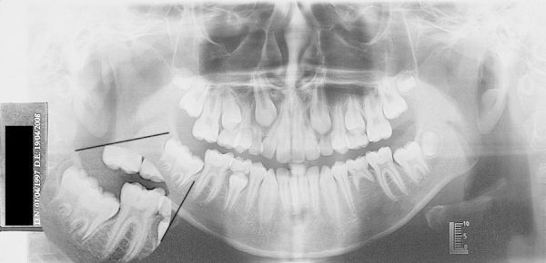
Panoramic Radiography (PR) for initial orthodontic treatment, showed second premolar agenesis (mandibular left region—35) and a second molar (mandibular right) apparently overlaid on the third molar, see that this tooth has more advanced root development than their congeners.
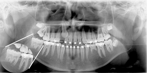
Second radiography, three years later, showed a second molar (mandibular right) had two separate canals and shared one (second molar mesial root and first molar distal root).

Intraoral photography. Clinically patient presented separated crowns and the number of teeth does not change.
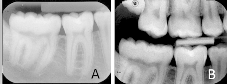
(A) Periapical radiography; (B) Bite-wing radiography.
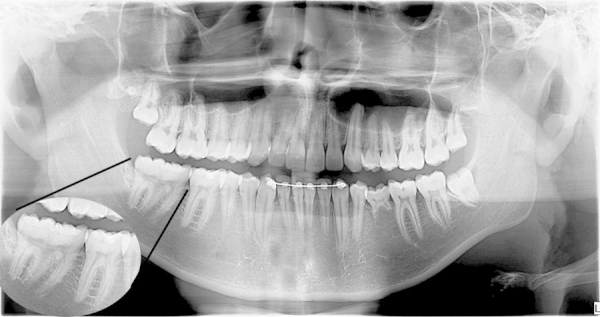
Third radiography presented a tooth with three roots, two crowns sharing a pulp chamber and one root canal (the other third molars are with open apex). Presence of superior forth molar.
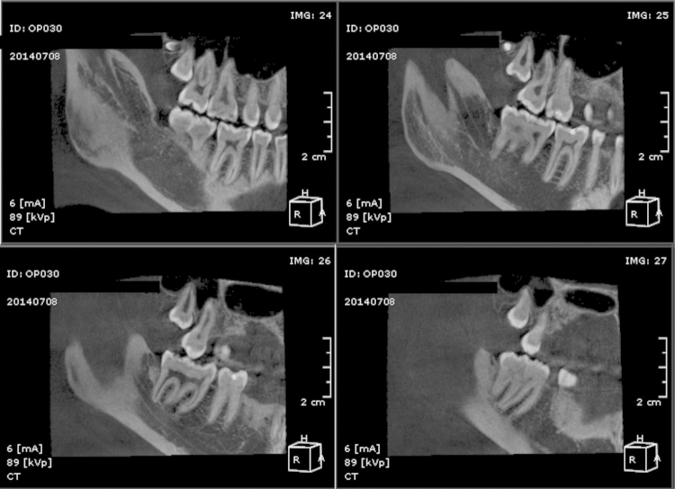
CT scan performed on CBCT (OP300-Instrumentarium Dental). Sagittal view, IMG: 24, the tooth in question due overlapping teeth presents enamel layer between the pulp chambers, observe the tooth 46 with central image, which indicates that the anomalous teeth is slightly off the dental arch. Note the superior forth molar presence. In the next slice (IMG: 25) presents clear union crown, showing a highlighted sulcus dividing the crowns. There caries lesion in the distal sulcus of the mesial crown. Clear presence of a “double teeth” (slice IMG: 26) with one pulp chamber, three root canals, mesial, distal and central. Note the pronounced pulp horn in the mesial which may indicate that it is possible a gemination.
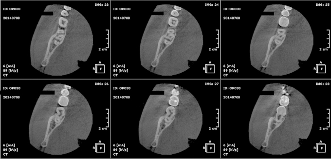
Axial view; the sequence showed initially three separate roots (IMG: 23 and 24), the next slice begins “fusion” of the mesial root with the “median” root. Slices IMG: 26 and IMG: 27, the pulp chamber is shared (the junction of the pulp chamber or the pulp portion of these roots), and finally showed the union, the mesial and median root are covered by dentin and is seen the distal root canal.
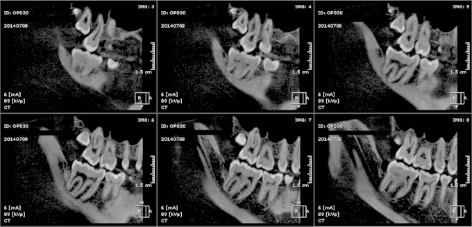
Panorama view; showed the same details of sagittal view. Note the curvature of mesial and distal roots, as if there were no central root.
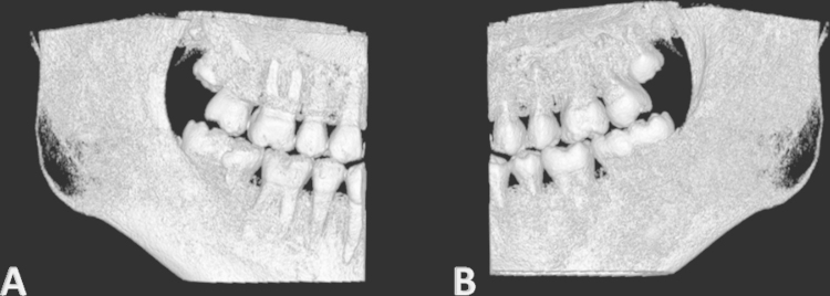
The 3D reconstruction. (A) Vestibular view. (B) Lingual view. 3D reconstruction corroborates the clinical vision.
References
-
- Grover P.S., Lorton L. Gemination and twinning in the permanent dentition. Oral Surg. Oral Med. Oral Pathol. 1985;59:313–318. - PubMed
-
- Kremeier K., Pontius O., Klaiber B., Hülsmann M. Nonsurgical endodontic management of a double tooth: a case report. Int. Endod. J. 2007;40:908–915. - PubMed
-
- Tannenbaum K.A., Ailing E.E. Anomalous tooth development: case reports of gemination and twinning. Oral Surg. Oral Med. Oral Pathol. 1963;16:883–887. - PubMed
-
- Stabholz A., Friedman F. Endodontic therapy of an unusual maxillary permanent first molar. J. Endod. 1983;9(7):293–295. - PubMed
LinkOut - more resources
Full Text Sources
Other Literature Sources
