A giant mucinous cystadenocarcinoma of the appendix: a case report and review of the literature
- PMID: 26945579
- PMCID: PMC4779566
- DOI: 10.1186/s12957-016-0828-2
A giant mucinous cystadenocarcinoma of the appendix: a case report and review of the literature
Abstract
Background: Mucinous cystadenocarcinoma is the second most common etiology of appendiceal mucocele. We report a relatively rare case of a giant appendiceal mucocele caused by mucinous cystadenocarcinoma, which occupied the entire abdomen of an adult woman.
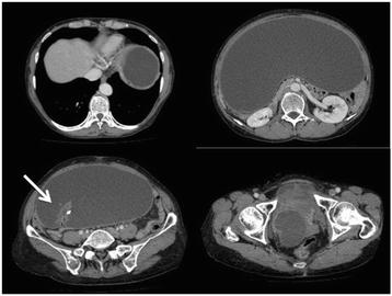
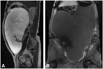
Case presentation: A 63-year-old woman presented with a chief complaint of abdominal distention. Imaging studies showed a giant cystic mass occupying her entire abdomen. Laparotomy confirmed a giant appendiceal mucocele, and the patient underwent ileocecal resection. A mucinous deposit was not found in her abdominal cavity, and the ovaries were grossly normal bilaterally. The pathological diagnosis was mucinous adenocarcinoma with a low-grade mucinous neoplasm that invaded the subserosa. Regional lymph node metastasis was not found. She has had recurrence-free survival for 5 years.
Conclusions: The present case is the largest appendiceal cystadenocarcinoma ever reported. The optimal treatment of an appendiceal neoplasm requires further research based on consensus terminology of an appendiceal mucocele.
Figures


Similar articles
-
[A case of mucinous cystadenocarcinoma of the appendix penetrating the urinary bladder].Hinyokika Kiyo. 2002 Jun;48(6):351-4. Hinyokika Kiyo. 2002. PMID: 12166235 Review. Japanese.
-
[Mucocele of the appendix - a heterogenous surgical pathology].Zentralbl Chir. 2010 Aug;135(4):330-5. doi: 10.1055/s-0029-1224563. Epub 2009 Dec 7. Zentralbl Chir. 2010. PMID: 19998220 German.
-
Low-grade appendiceal mucinous neoplasm and endometriosis of the appendix.World J Surg Oncol. 2017 Dec 19;15(1):226. doi: 10.1186/s12957-017-1294-1. World J Surg Oncol. 2017. PMID: 29258523 Free PMC article.
-
Heterotopic ossification in appendiceal mucinous neoplasms: clinicopathological characteristics of 3 cases.Malays J Pathol. 2016 Apr;38(1):49-54. Malays J Pathol. 2016. PMID: 27126665
-
A case report of a giant appendiceal mucocele and literature review.Pan Afr Med J. 2017 Oct 4;28:106. doi: 10.11604/pamj.2017.28.106.13832. eCollection 2017. Pan Afr Med J. 2017. PMID: 29515724 Free PMC article. Review.
Cited by
-
Challenges in diagnosing pseudomyxoma peritonei in a hepatitis B patient: A case report.Medicine (Baltimore). 2025 Apr 25;104(17):e42298. doi: 10.1097/MD.0000000000042298. Medicine (Baltimore). 2025. PMID: 40295254 Free PMC article.
-
A rare case of a large low-grade appendicular mucinous neoplasm causing compressive symptoms.J Surg Case Rep. 2022 Apr 13;2022(4):rjac111. doi: 10.1093/jscr/rjac111. eCollection 2022 Apr. J Surg Case Rep. 2022. PMID: 35432918 Free PMC article.
-
Ileocecal mucinous carcinoma misdiagnosed as incarcerated hernia: A case report.Open Life Sci. 2024 Dec 16;19(1):20221002. doi: 10.1515/biol-2022-1002. eCollection 2024. Open Life Sci. 2024. PMID: 39711975 Free PMC article.
-
Endoscopic diagnosis and treatment of an appendiceal mucocele: A case report.World J Clin Cases. 2021 Jun 6;9(16):3936-3942. doi: 10.12998/wjcc.v9.i16.3936. World J Clin Cases. 2021. PMID: 34141750 Free PMC article.
-
Primary adenocarcinoma of the appendix in a child: A case report.Surg Case Rep. 2018 Sep 4;4(1):109. doi: 10.1186/s40792-018-0514-4. Surg Case Rep. 2018. PMID: 30182291 Free PMC article.
References
-
- Rokitansky CF. A manual of pathological anatomy. Philadelphia: Blanchard & Lea; 1855.
Publication types
MeSH terms
LinkOut - more resources
Full Text Sources
Other Literature Sources

