Review
. 2016 May 25;583(1):64-77.
doi: 10.1016/j.gene.2016.02.042. Epub 2016 Mar 3.

Structure and function of the AAA+ ATPase p97/Cdc48p

Affiliations
Review

Structure and function of the AAA+ ATPase p97/Cdc48p

Di Xia et al. Gene. .

Abstract

p97 (also known as valosin-containing protein (VCP) in mammals or Cdc48p in Saccharomyces cerevisiae) is an evolutionarily conserved ATPase present in all eukaryotes and archaebacteria. In conjunction with a collection of cofactors and adaptors, p97/Cdc48p performs an array of biological functions mostly through modulating the stability of 'client' proteins. Using energy from ATP hydrolysis, p97/Cdc48p segregates these molecules from immobile cellular structures such as protein assemblies, membrane organelles, and chromatin. Consequently, the released polypeptides can be efficiently degraded by the ubiquitin proteasome system or recycled. This review summarizes our current understanding of the structure and function of this essential cellular chaperoning system.

Keywords: AAA+ ATPase; Chromatin-associated degradation; ER-associated degradation; Membrane fusion; Mitochondria-associated degradation; Proteasome; Protein quality control; Ribosome-associated degradation; Segregase; Ubiquitin; Unfoldase; p97/CDC48.


Figures

Figure 1
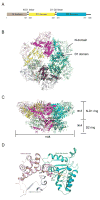
Figure 1. Structure of p97/Cdc48p
(A) The schematic domain organization of p97/Cdc48. (B) The structure of hexameric p97 (PDB: 3CF2 in the ADP-bound form) is viewed down the 6-fold symmetry axis showing the N-D1 ring. The six subunits are shown as cartoon diagrams in different colors. Domains of each subunit are also shaded differently. The D1 domain and the N-domain are indicated with arrows and labeled for one of the six subunits. (C) The side view of p97 is presented with indicated width and height. (D) The structure of the D1 AAA domain of a p97 subunit with bound ATPγS is presented in the ribbon format (PDB:4KO8). An AAA domain consists of a RecA-like domain (cyan) and a characteristic helical domain (purple). An ATPγS, bound at the interface between the two domains, is shown as stick model. The Mg2+ ion and three conserved water molecules are shown as silver and red balls, respectively. The Walker A motif or P-loop is highlighted in red and the conserved lysine residue K251 is shown as stick model and labeled. The Walker B motif is shown in orange and the two conserved acidic residues D304 and E305 are represented by stick models. The nucleotide-binding site communicates with a neighboring subunit through the SRH (second region of homology, in light blue) motif, where a conserved Arg-finger residue R359 is in contact with the bound nucleotide.
Figure 2
Figure 2. Nucleotide-dependent N-domain conformational change
A large N-domain conformational change has been observed, being driven by the nucleotide cycle of the D1 domain. When ATP is bound, the N-domain, illustrated in ribbon diagram in magenta, goes to the Up-conformation, whereas it moves to the Down-conformation when ADP is bound to the D1 domain. During the transition between the UP- and Down-conformation, the center of gravity of the N-domain (B for the Down-conformation and C for the Up-conformation) translated by 13 Å and the α angle is 11° as defined by the triangle ABC (A is the position of G208 of the D1 domain). Additionally, to adopt the Up-conformation configuration, the N-domain needs a further 92° rotation. As a result, the residue P178 moves up by 38 Å.
Figure 3
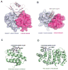
Figure 3. The interactions of p97 with adaptors and cofactors
(A) Structure of the p97 N-domain in complex with the UBX domain of FAF1 (PDB:3QC8). The N-domain, depicted as a molecular surface overlaid to a ribbon representation, consists of two subdomains: N-terminal double Ψ-barrel domain (purple) and C-terminal β-barrel domain (red). The UBX domain of FAF1 is depicted as ribbon diagram in magenta. Critical residues for interaction are shown as ball-and-stick models and labeled. (B) Structure of the p97 N-domain in complex with the VIM motif of gp78 (PDB:3TIW). Here the VIM motif is shown as helix in brown and its binding to the N-domain is mostly mediated by charged residues. (C) Structure of the N-terminal domain of PNGase in complex with a C-terminal peptide of p97 (PDB:2HPL). The PNGase N-terminal domain is shown in cartoon representation in green. The bound peptide is shown as a stick model with five residues (labeled) seen in the structure. The carbon atoms are colored in black, nitrogen in blue and oxygen in red. (D) Structure of the PUL domain of FLAA/Ufd3 in complex with a C-terminal peptide of p97 (PDB:3EBB). The PLAA PUL domain is shown in cartoon representation in green. The bound peptide is shown as a stick model with four residues visible in the structure. The carbon atoms are colored in black, nitrogen in blue and oxygen in red.
Figure 4
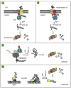
Figure 4. The established segregase function of p97/Cdc48p
p97 collaborates with the proteasome in degradation of misfolded ER proteins (the ERAD pathway) (A), misfolded proteins in the mitochondrial outer membrane (B), defective translocation products (C), and chromatin-associated proteins (D). In each scenario, p97 uses energy from ATP hydrolysis to release polypeptides from either the membranes, the ribosome, or DNA, and then target them to the proteasome for degradation. E3, ubiquitin ligase, R, retrotranslocation complex, mitochondrial IS, mitochondrial inter-membrane space.
Figure 5
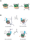
Figure 5. Force generation coupled to ATP hydrolysis
(A) An ATP hydrolysis model for p97. A p97 hexamer is represented as two concentric rings with D1 ring in green and D2 ring in brown. The N-domains in the Down-conformation are shown as magenta balls. D1 domains with pre-bound ADP are labeled with the letter D. ATP molecules introduced into the system will first go to the D1 domains with no pre-bound nucleotide, which leads the N-domains to the Up-conformation. Occupation of ATP to the D1 domain renders the cognate D2 domain capable of hydrolyzing ATP, which is labeled with a red *. The D1 domain probably hydrolyzes ATP once a few D2 domains have been converted to the ADP bound state. (B) A proposed model of force generation by a N-domain conformational change in ERAD. p97 is anchored to the ER membrane by association with a membrane adaptor (blue) using its N-domain. If substrate is bound to the central pore of the D2 domain when the D1 domain is in the Up-conformation (top panel), the swing of the N-domain to the Down-conformation after ATP hydrolysis will pull the D1 ring closer to the membrane, leading to the extraction of substrate out of the membranes. Alternatively, if the substrate is bound to the D1 domain (bottom panel), a switch from the Down-conformation to the Up-conformation following nucleotide exchange in D1 will move the ATPase domains away from the membrane, pulling substrate out of the membrane.

References

    1. Acharya U, Jacobs R, Peters JM, Watson N, Farquhar MG, Malhotra V. The formation of Golgi stacks from vesiculated Golgi membranes requires two distinct fusion events. Cell. 1995;82:895–904. - PubMed
    1. Acs K, Luijsterburg MS, Ackermann L, Salomons FA, Hoppe T, Dantuma NP. The AAA-ATPase VCP/p97 promotes 53BP1 recruitment by removing L3MBTL1 from DNA double-strand breaks. Nature structural & molecular biology. 2011;18:1345–1350. - PubMed
    1. Allen MD, Buchberger A, Bycroft M. The PUB domain functions as a p97 binding module in human peptide N-glycanase. The Journal of biological chemistry. 2006;281:25502–25508. - PubMed
    1. Anderson DJ, Le Moigne R, Djakovic S, Kumar B, Rice J, Wong S, Wang J, Yao B, Valle E, Kiss von Soly S, Madriaga A, Soriano F, Menon MK, Wu ZY, Kampmann M, Chen Y, Weissman JS, Aftab BT, Yakes FM, Shawver L, Zhou HJ, Wustrow D, Rolfe M. Targeting the AAA ATPase p97 as an Approach to Treat Cancer through Disruption of Protein Homeostasis. Cancer cell. 2015;28:653–665. - PMC - PubMed
    1. Ballar P, Shen Y, Yang H, Fang S. The role of a novel p97/valosin-containing protein-interacting motif of gp78 in endoplasmic reticulum-associated degradation. The Journal of biological chemistry. 2006;281:35359–35368. - PubMed

Publication types

MeSH terms

LinkOut - more resources