. 2016 Apr 15;412(2):208-18.
doi: 10.1016/j.ydbio.2016.01.042. Epub 2016 Mar 3.

Coiled-coil domain containing 42 (Ccdc42) is necessary for proper sperm development and male fertility in the mouse

Affiliations

Coiled-coil domain containing 42 (Ccdc42) is necessary for proper sperm development and male fertility in the mouse

Raymond C Pasek et al. Dev Biol. .

Abstract

Spermiogenesis is the differentiation of spermatids into motile sperm consisting of a head and a tail. The head harbors a condensed elongated nucleus partially covered by the acrosome-acroplaxome complex. Defects in the acrosome-acroplaxome complex are associated with abnormalities in sperm head shaping. The head-tail coupling apparatus (HTCA), a complex structure consisting of two cylindrical microtubule-based centrioles and associated components, connects the tail or flagellum to the sperm head. Defects in the development of the HTCA cause sperm decapitation and disrupt sperm motility, two major contributors to male infertility. Here, we provide data indicating that mutations in the gene Coiled-coil domain containing 42 (Ccdc42) is associated with malformation of the mouse sperm flagella. In contrast to many other flagella and motile cilia genes, Ccdc42 expression is only observed in the brain and developing sperm. Male mice homozygous for a loss-of-function Ccdc42 allele (Ccdc42(KO)) display defects in the number and location of the HTCA, lack flagellated sperm, and are sterile. The testes enriched expression of Ccdc42 and lack of other phenotypes in mutant mice make it an ideal candidate for screening cases of azoospermia in humans.

Keywords: Ccdc42; Infertility; Intramanchette transport; Sperm; Sperm head–tail coupling apparatus.


Figures

Figure 1
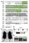
Figure 1. Ccdc42 homologs and generation of a knockout mouse allele
(A) ClustalW protein alignment of the human, mouse, Tetrahymena thermophila, and Chlamydomonas reinhardtii Ccdc42 homologs. Below the alignment (*) indicates identical, (:) conserved, and (.) semi-conservative amino acids. Gray highlight indicates coiled-coil motif. Green highlight indicates DUF4200 domains. (B) The Ccdc42 knockout allele was generated by replacing all 7 exons with a lacZ-p(A) reporter cassette (blue box). Location of primers used for genotyping are indicated by the blue and green triangles. (C) Wild-type control (Ccdc42WT) mouse next to a knockout (Ccdc42KO) sibling show no overt phenotypes in the mutants. (D) Genomic DNA PCR genotyping of wild-type (Ccdc42WT), heterozygous (Ccdc42Het) and knockout (Ccdc42KO) mice.
Figure 2
Figure 2. Ccdc42 expression in the mouse
(A) RT-PCR for Ccdc42 in several tissues derived from 8-week old mice. Expression is limited to testes and brain. (B) RT-PCR analysis for Ccdc42 in the developing testes. Expression begins at approximately 10 days of age. Brain Ccdc42 expression was present in 8 day old mice prior to testes expression. (A and B) β-Actin is used as a template control. (C) β-galactosidase staining assay for Ccdc42-β-gal reporter in the testes and brain. β-galactosidase activity is present in maturing spermatids in the adluminal region of the seminiferous tubule of Ccdc42Het testes. Ccdc42KO sections reveal staining in malformed spermatids. Ccdc42WT testes are a negative control for staining. Purkinje cells of the cerebellum stain positive for β-galactosidase activity. Sections counterstained with Fast Red. All scale bars 60 μm.
Figure 3
Figure 3. Testicular and epididymal phenotypes in Ccdc42KO mice
(A) Periodic acid-Schiff and hematoxylin staining of testes cross sections from 8 week old control (Ccdc42WT) and mutant (Ccdc42KO) mice. Ccdc42KO mice display both an absence of a lumen and spermatid flagella. Scale bars 60 μm. (C) High magnification plastic section through a seminiferous tubule stained with toluidine blue. Mature spermatids are released into the lumen during spermiation in Ccdc42WT mice. (D) Spermiation stage in the seminiferous tubule of Ccdc42KO mice. Arrows indicate mature spermatids with abnormal shaped heads. No flagella tails are observed in Ccdc42KO. (E) Epididymal duct in a Ccdc42WT mouse with flagellated sperm. (F) Epididymis from an age matched Ccdc42KO mouse devoid of sperm contains only cellular debris. (C–F) Scale bars 5 μm.
Figure 4
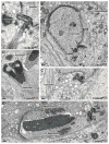
Figure 4. Morphological defects in Ccdc42KO spermatids
(A–E) Transmission electron micrographs of spermatids. (A) The HTCA in an elongating spermatid of a Ccdc42WT mouse. The following are indicated: (1) nuclear implantation fossa, (2) capitulum, a dense plate facing the implantation fossa, (3) proximal centriole, (4) distal centriole, the emerging site of the, (6) axoneme, (5) banded collar surrounding the HTCA, A typical chromatoid body and adjacent vesicles are also visible. (B) Elongating spermatid of a Ccdc42KO mouse. Three HTCAs (arrows) are present in this section, surrounded by numerous particle-filled vesicles. Two HTCAs are anchored to implantation fossae, the other HTCA is separate from the nucleus. The microtubules of the manchette are present, but no microtubule axonemes are observed from any of the HTCAs. The marginal ring of the acroplaxome (Mr apx) and the associated acrosome are present but mitochondria are dispersed. There is no indication of outer dense fiber assembly. (C) A structurally normal looking HTCA from a mutant mouse is dissociated from its anchoring site and has no axoneme. The microtubules of the manchette and adjacent vesicles in the elongating spermatid of a Ccdc42KO mouse appear normal. (D) A Ccdc42KO condensed spermatid nucleus with abnormal nuclear folding. The manchette is still present. (E) A nearly mature spermatid in the Ccdc42KO with two HTCAs (arrows) attached to a fragment of nucleus (*). There is no evidence of flagellogenesis. The nuclear envelope (Nucl Env) is detached from the condensed chromatin and clusters of mitochondria can be seen.
Figure 5
Figure 5. GFP-Ccdc42 localizes to the base of cilia in transgenic Tetrahymena thermophila
(A) In the uninduced state, no GFP signal is detected in transgenic GFP-Ccdc42 cells. Cilia axonemes (red) have been labeled with a polyglycylated tubulin (2302) antibody. Cilia tips (white) have been labeled with an α-tubulin antibody (12G10). (B) Two hours after the addition of CdCl2 to induce transgene expression, GFP-Ccdc42 (green) can be seen at the bases of the cilia. Nuclei have been labeled with Hoechst. Scale bar is 30 μm. Higher magnification images are shown in the bottom panels.

References

    1. Blanchon S, Legendre M, Copin B, Duquesnoy P, Montantin G, Kott E, Dastot F, Jeanson L, Cachanado M, Rousseau A, Papon JF, Beydon N, Brouard J, Crestani B, Deschildre A, Desir J, Dollfus H, Leheup B, Tamalet A, Thumerelle C, Vojtek AM, Escalier D, Coste A, de Blic J, Clement A, Escudier E, Amselem S. Delineation of CCDC39/CCDC40 mutation spectrum and associated phenotypes in primary ciliary dyskinesia. J Med Genet. 2012;49:410–416. - PubMed
    1. Borg CL, Wolski KM, Gibbs GM, O'Bryan MK. Phenotyping male infertility in the mouse: how to get the most out of a 'non-performer'. Human reproduction update. 2010;16:205–224. - PMC - PubMed
    1. Buffone MG, Ijiri TW, Cao W, Merdiushev T, Aghajanian HK, Gerton GL. Heads or tails? Structural events and molecular mechanisms that promote mammalian sperm acrosomal exocytosis and motility. Molecular reproduction and development. 2012;79:4–18. - PMC - PubMed
    1. Chizhikov VV, Davenport J, Zhang Q, Shih EK, Cabello OA, Fuchs JL, Yoder BK, Millen KJ. Cilia proteins control cerebellar morphogenesis by promoting expansion of the granule progenitor pool. The Journal of neuroscience : the official journal of the Society for Neuroscience. 2007;27:9780–9789. - PMC - PubMed
    1. De Kretser DM, Baker HW. Infertility in men: recent advances and continuing controversies. The Journal of clinical endocrinology and metabolism. 1999;84:3443–3450. - PubMed

Publication types

MeSH terms