. 2016 Mar 7:7:10866.
doi: 10.1038/ncomms10866.

CSF-contacting neurons regulate locomotion by relaying mechanical stimuli to spinal circuits

Affiliations

CSF-contacting neurons regulate locomotion by relaying mechanical stimuli to spinal circuits

Urs Lucas Böhm et al. Nat Commun. .

Abstract

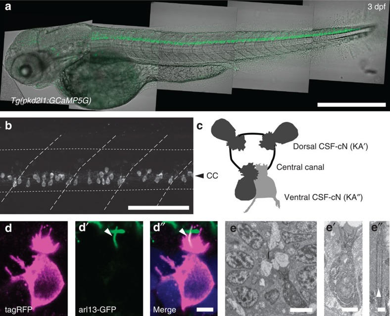
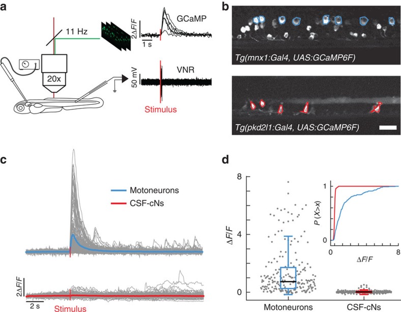
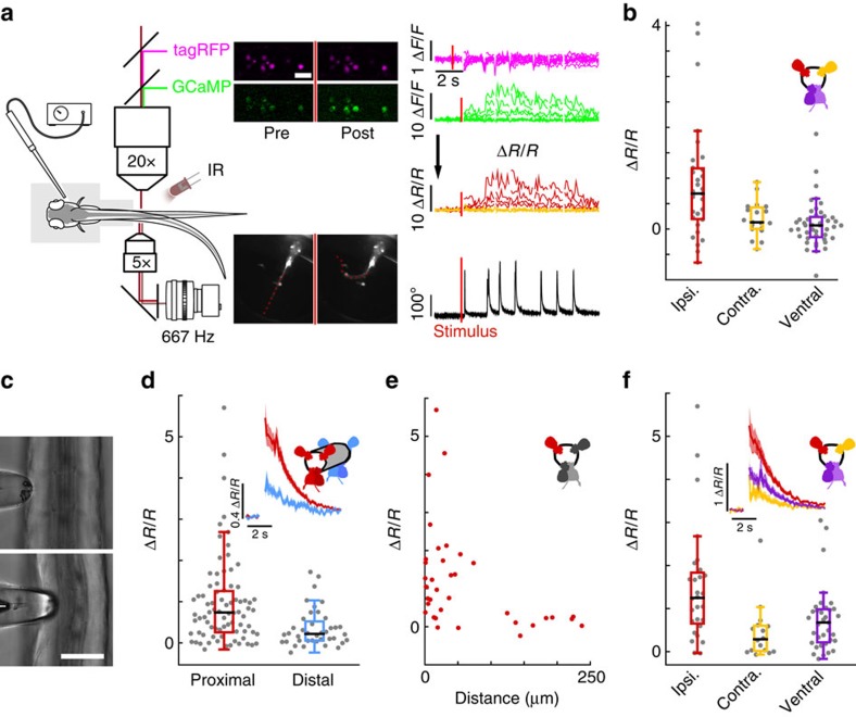
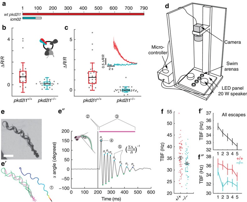
Throughout vertebrates, cerebrospinal fluid-contacting neurons (CSF-cNs) are ciliated cells surrounding the central canal in the ventral spinal cord. Their contribution to modulate locomotion remains undetermined. Recently, we have shown CSF-cNs modulate locomotion by directly projecting onto the locomotor central pattern generators (CPGs), but the sensory modality these cells convey to spinal circuits and their relevance to innate locomotion remain elusive. Here, we demonstrate in vivo that CSF-cNs form an intraspinal mechanosensory organ that detects spinal bending. By performing calcium imaging in moving animals, we show that CSF-cNs respond to both passive and active bending of the spinal cord. In mutants for the channel Pkd2l1, CSF-cNs lose their response to bending and animals show a selective reduction of tail beat frequency, confirming the central role of this feedback loop for optimizing locomotion. Altogether, our study reveals that CSF-cNs constitute a mechanosensory organ operating during locomotion to modulate spinal CPGs.


Figures

Figure 1
Figure 1. In the spinal cord, pkd2l1+CSF-cNs project an apical extension consisting of one kinocilium and a brush of microvilli into the central canal.
(a) Transmitted and fluorescent image showing that the pkd2l1 promoter in Tg(pkd2l1:GCaMP5G) transgenic larva drives GCaMP5G expression in CSF-cNs along the entire spinal cord. Scale bar: 500 μm. (b) A close up from a lateral view in the same transgenic animal shows the morphology of CSF-cNs as elongated cells with an apical extension reaching the central canal. Scale bar: 100 μm. (c) Schematic depicting dorsal and ventral CSF-cN location around the central canal. (d) Confocal microscopy of 50 μm sections of the 4 dpf triple transgenic larva Tg(pkd2l1:gal4, UAS:tagRFP-CAAX;cmcl2:GFP, βact:Arl13-GFP) shows that CSF-cNs project one Arl13-GFP+ cilium (arrowhead) and multiple microvilli into the CSF. Scale bar: 3 μm. (e) Transmission electron microscopy in Tg(pkd2l1:Gal4) larvae injected with UAS:APEX2-tagRFP shows a single ventral CSF-cN reaching the central canal (close up in (e′), scale bar: 2 μm) and exhibiting a motile 9+2 cilium (arrowhead, (e″), scale bar: 250 nm).
Figure 2
Figure 2. CSF-cNs are minimally activated during fictive escapes when no muscle contraction occurs.
(a) Schematic view of the experimental setup combining 2-photon laser scanning microscope for calcium imaging and electrophysiological recordings of the ventral nerve root. 10 ms-long water jets delivered to the otic vesicle triggered fictive escapes in paralyzed larvae. (b) Lateral view showing expression of GCaMP6f in MNs in the double transgenic larva Tg(mnx1:Gal4, UAS:GCaMP6f;cryaa:mCherry) and in CSF-cNs in Tg(pkd2l1:gal4, UAS:GCaMP6f;cryaa:mCherry). ROIs indicate cells included in the analysis. Only the dorsalmost MNs were analyzed in the mnx1 line. Scale bar: 20 μm. (c) Typical calcium transients recorded in MNs (blue) and in CSF-cNs (red) during fictive escapes; ‘stimulus' indicates when the water jet was triggered, average response in coloured lines. (d) Quantification of calcium transient amplitude in MNs and CSF-cNs (each data point represents one recording from one cell; plots use median as the measure of central tendency; inset is the cumulative histogram of calcium responses). Responses in both populations are greater than baseline (204 MNs from 6 larvae: mean ΔF/F=1.2, P<1.0 × 10⁻⁸; 192 CSF-cNs from 7 larvae: mean ΔF/F=0.042, P=3.78 × 10⁻⁸), but CSF-cNs exhibit significantly less activity than MNs (P=0.014).
Figure 3
Figure 3. CSF-cNs respond to active muscle contraction as well as to passive mechanical bending of the spinal cord.
(a) Schematic describing 2-photon imaging experiments used to record simultaneously from CSF-cNs expressing tagRFP (magenta) and GCaMP5 (green) in head-embedded Tg(pkd2l1:GCaMP5, pkd2l1:tagRFP) larvae. Infrared illumination combined with high-speed video recording shows unidirectional tail deflections induced by a water jet to the otic vesicle. Sample traces for ΔF/F of tagRFP and GCaMP shown with tail deflection during escape (note: vertical scale is 10 times larger for GCaMP compared to tagRFP signals). Subtracting the tagRFP signal from the GCaMP signal removed motion artifacts; breaks in the trace arise from frames when cells escaped from the focal plane. Scale bar: 10 μm. (b) Quantification of calcium transient amplitude in response to muscle contraction (n=11 larvae) in dorsal CSF-cNs either ipsilateral (red, 31 cells) or contralateral (yellow, 19 cells) or ventral (purple, 44 cells). Only dorsal ipsilateral cells exhibited responses greater than baseline (P=9.51 × 10⁻⁴) and all other cell types responded significantly less than dorsal ipsilateral cells (dorsal contralateral: P=2.43 × 10⁻³, ventral: P=4.85 × 10⁻⁵). (c) Passive mechanical stimulation of CSF-cNs in paralyzed larvae (n=5) was implemented with mechanical pressure exerted by pushing a glass probe laterally against the fish tail. Scale bar: 50 μm. (d) Response of proximal (<100 μm) and distal (>100 μm) CSF-cNs. Inset: average calcium response of proximal (red) versus distal (blue) CSF-cNs. (e) Response of dorsal ipsilateral (red) CSF-cNs as function of distance from the probe. (f) Response of dorsal ispsilateral (red, 28 cells), dorsal contralateral (yellow, 16 cells) and ventral (purple, 36 cells) CSF-cNs relative to the location of mechanical stimulation. Inset: Average calcium response of dorsal ispsilateral versus dorsal contralateral and ventral CSF-cNs. All cell types show a response different from 0 (dorsal ipsilateral: P<1.0 × 10⁻⁸, dorsal contralateral: P=5.95 × 10⁻⁵, ventral: P<1.0 × 10⁻⁸) and all other cell types responded significantly less than dorsal ipsilateral cells (dorsal contralateral: P=1.06 × 10⁻³, ventral: P=7.22 × 10⁻³).
Figure 4
Figure 4. Mutation of pkd2l1 abolishes CSF-cN response to active and passive spinal bending and leads to a reduction of swimming frequency during the escape.
(a) The TALEN-mediated 8 nucleotide deletion in exon two of pkd2l1 leads to a frame shift and a premature stop in pkd2l1icm02. (b,c) Calcium transients in dorsal ipsilateral CSF-cNs are abolished in pkd2l1 mutant after mechanical stimulation during active (b) and passive (c) tail movement (same stimulation paradigms as used in Fig. 3, wildtype data sets for (b,c) are the same as in Fig. 3). Mutants (10 fish, 29 cells) in b show no response different from 0 (P=0.13) and are different from wildtypes (11 fish, 31 cells, P=1.84 × 10⁻²). Mutants (7 fish, 30 cells) in c show no response different from 0 (P=0.22) and are different from wildtypes (5 fish, 28 cells, P=2.79 × 10⁻⁴). (d) Experimental setup monitoring at high speed the escape response of freely swimming zebrafish larvae isolated in separated swim arenas triggered by 10 ms, 500 Hz acoustic stimuli. Acoustic stimuli were repeated five times per larva with 2 min inter trial interval. (e) Superimposed images showing a typical acoustic escape, (e′) tracking corresponding to the same escape, swim distance (1) is derived from swim bladder position. (e″) kinematic analysis relied on the measure of the tail angle over time α(t) enabling to measure latency (2), escape duration (3), C-bend amplitude (4) and number of oscillations and TBF based on detection of subsequent peaks (5). Speed is derived from swim distance divided by duration. Scale bar: 2 mm. (f) Reduction of TBF in the pkd2l1 mutant. Each point corresponds to a value per larva averaged over multiple trials (P=0.0091). (f′) TBF decreased across trials in our paradigm (P=1.7 × 10⁻⁵). (f″) The reduction of TBF in the pkd2l1 mutant (blue) compared to wild type is noticeable across trials (P=0.039).

References

    1. Martin F. H., Seoane J. R. & Baile C. A. Feeding in satiated sheep elicited by intraventricular injections of CSF from fasted sheep. Life Sci. 13, 177–184 (1973). - PubMed
    1. Pappenheimer J. R., Miller T. B. & Goodrich C. A. Sleep-promoting effects of cerebrospinal fluid from sleep-deprived goats. Proc. Natl Acad. Sci. USA 58, 513–517 (1967). - PMC - PubMed
    1. Lehtinen M. K. et al. The cerebrospinal fluid provides a proliferative niche for neural progenitor cells. Neuron 69, 893–905 (2011). - PMC - PubMed
    1. Sawamoto K. et al. New neurons follow the flow of cerebrospinal fluid in the adult brain. Science 311, 629–632 (2006). - PubMed
    1. Xie L. et al. Sleep drives metabolite clearance from the adult brain. Science 342, 373–377 (2013). - PMC - PubMed

Publication types

MeSH terms

Substances

LinkOut - more resources