Review
. 2016 Aug;1857(8):1219-1227.
doi: 10.1016/j.bbabio.2016.02.020. Epub 2016 Mar 4.

Role of cysteines in mammalian VDAC isoforms' function

Affiliations
Review

Role of cysteines in mammalian VDAC isoforms' function

Vito De Pinto et al. Biochim Biophys Acta. 2016 Aug.

Abstract

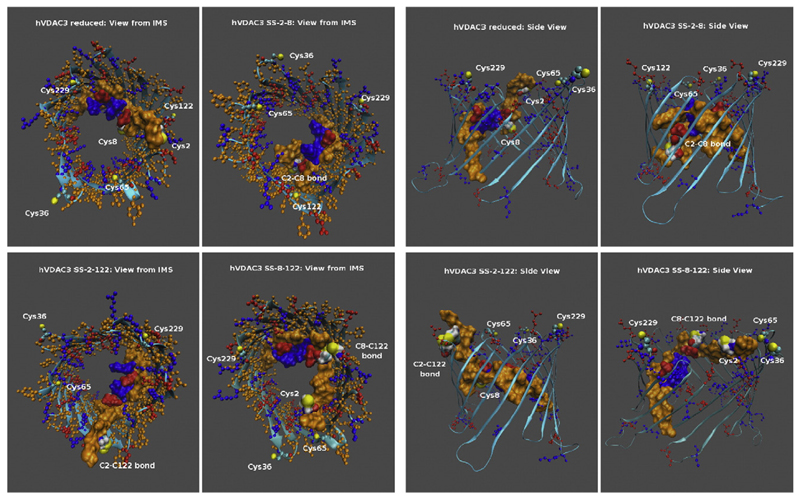
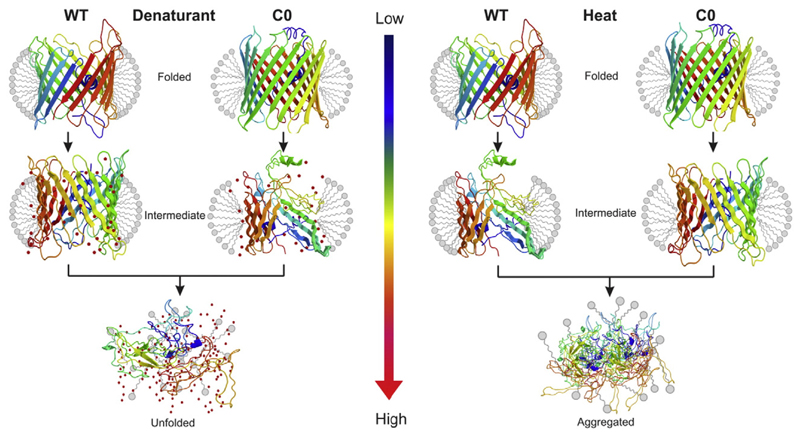
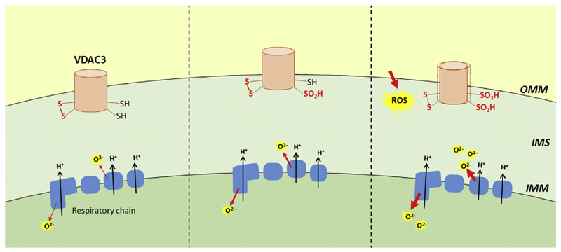
In this mini-review, we analyze the influence of cysteines in the structure and activity of mitochondrial outer membrane mammalian VDAC isoforms. The three VDAC isoforms show conserved sequences, similar structures and the same gene organization. The meaning of three proteins encoded in different chromosomes must thus be searched for subtle differences at the amino acid level. Among others, cysteine content is noticeable. In humans, VDAC1 has 2, VDAC2 has 9 and VDAC3 has 6 cysteines. Recent works have shown that, at variance from VDAC1, VDAC2 and VDAC3 exhibit cysteines predicted to protrude towards the intermembrane space, making them a preferred target for oxidation by ROS. Mass spectrometry in VDAC3 revealed that a disulfide bridge can be formed and other cysteine oxidations are also detectable. Both VDAC2 and VDAC3 cysteines were mutagenized to highlight their role in vitro and in complementation assays in Δporin1 yeast. Chemico-physical techniques revealed an important function of cysteines in the structural stabilization of the pore. In conclusion, the works available on VDAC cysteines support the notion that the three proteins are paralogs with a similar pore-function and slightly different, but important, ancillary biological functions. This article is part of a Special Issue entitled 'EBEC 2016: 19th European Bioenergetics Conference, Riva del Garda, Italy, July 2-6, 2016', edited by Prof. Paolo Bernardi.


Figures

Fig. 1
Fig. 1
Predicted structures of the hVDAC3 channel in the reduced state and of the same protein containing respectively disulfide SS-2–8, SS-2–122 and SS-8–122. Left Panel. The figures show a view of the pores from the intermembrane mitochondrial space (IMS). The N-terminal tail (residues 1–25) containing cysteines 2 and 8, both involved in the predicted disulfide bridges, is shown in a space-filling representation to highlight its position with respect to the pore. Basic and acidic residues as well as cysteines are shown in a ball-and-stick representation. Basic residues are colored in blue while the acidic ones are in red and the neutral ones in orange. Sulfur atoms are colored in yellow. The structures were generated through a 400 ns MD simulation in the NVT ensemble as detailed in [60]. The Figure was produced using the VMD program [76]. Right Panel. The figures show a side-view of the pores. The same conventions and the same program were used in this panel. The authors are indebted to Carlo Guardiani (Cagliari, Warwick) for the figure drawing.
Fig. 2
Fig. 2
Schematic representation of the effect of denaturant on hVDAC2 WT and C0 mutants. Cartoon representation of the barrel (rainbow color) is shown in the folded conformation, and is surrounded by a detergent micelle (gray color). The colored arrow represents an increasing denaturant concentration (gradient increases from blue to red color). The left panel highlights the interaction of WT and C0 with a chemical denaturant, such as guanidine hydrochloride. Proteins remain folded when the denaturant is absent, but increasing the chemical denaturant results in unfolding of protein by disrupting protein–lipid interaction. As WT exhibits stronger protein–lipid interactions, the barrel retains considerable structure at intermediate denaturant concentrations, compared to the C0 protein. The right panel represents the response of hVDAC2 mutants to thermal denaturation. Here, increasing the temperature disrupts protein–protein and protein–lipid interaction, and finally gives rise to the formation of irreversible protein aggregates. C0 is well structured and forms stronger intra-protein contacts. Hence, these non-covalent interactions allow the barrel to remain structured at temperatures that cause WT unfolding.
Fig. 3
Fig. 3
Change in VDAC3 oxidation states due to elevated ROS level can lead to pore modifications. VDAC3 cysteines are in a stationary situation, most likely mainly reduced, and protruding towards the mitochondrial intermembrane space (left). Complex III and other proteins pour ROS in the IMS. The progressive accumulation of ROS increases the amount of oxidized cysteines in VDAC3. Also sulfinic and sulfonic states can be reached, randomly, in various exposed cysteines (central panel). Continuous increase of ROS in IMS can heavily modify VDAC3 cysteines and these irreversible modifications can be supposed to produce conformational changes with various possible outputs like definitive damaging of the protein or changes signaling the ROS level to surrounding actors (right panel). The oxidation state of cysteines to –SO2H and –SO3H is considered "irreversible" since neither of these oxoforms can be reduced directly by cellular thiols.

References

    1. Bachi A, Dalle-Donne I, Scaloni A. Redox proteomics: chemical principles, methodological approaches and biological/biomedical promises. Chem Rev. 2013;113:596–698. - PubMed
    1. Benz R. Permeation of hydrophilic solutes through mitochondrial outer membranes: review on mitochondrial porins. Biochim Biophys Acta. 1994;1197:167–196. - PubMed
    1. Colombini M, Blachly-Dyson E, Forte M. VDAC, a channel in the outer mitochondrial membrane. Ion Channels. 1996;4:169–202. - PubMed
    1. Shoshan-Barmatz V, De Pinto V, Zweckstetter M, Raviv Z, Keinan N, Arbel N. VDAC, a multi-functional mitochondrial protein regulating cell life and death. Mol Asp Med. 2010;31:227–285. - PubMed
    1. Cortese JD, Voglino AL, Hackenbrock CR. The ionic strength of the intermembrane space of intact mitochondria is not affected by the pH or volume of the intermembrane space. Biochim Biophys Acta. 1992;1100:189–197. - PubMed

Publication types

MeSH terms

Substances

LinkOut - more resources