. 2016 Mar 15;14(10):2301-12.
doi: 10.1016/j.celrep.2016.02.036. Epub 2016 Mar 3.

YAP Induces Human Naive Pluripotency

Affiliations

YAP Induces Human Naive Pluripotency

Han Qin et al. Cell Rep. .

Abstract

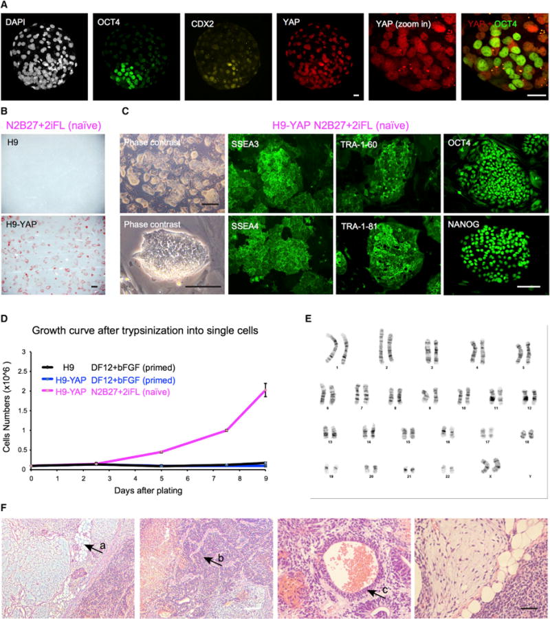
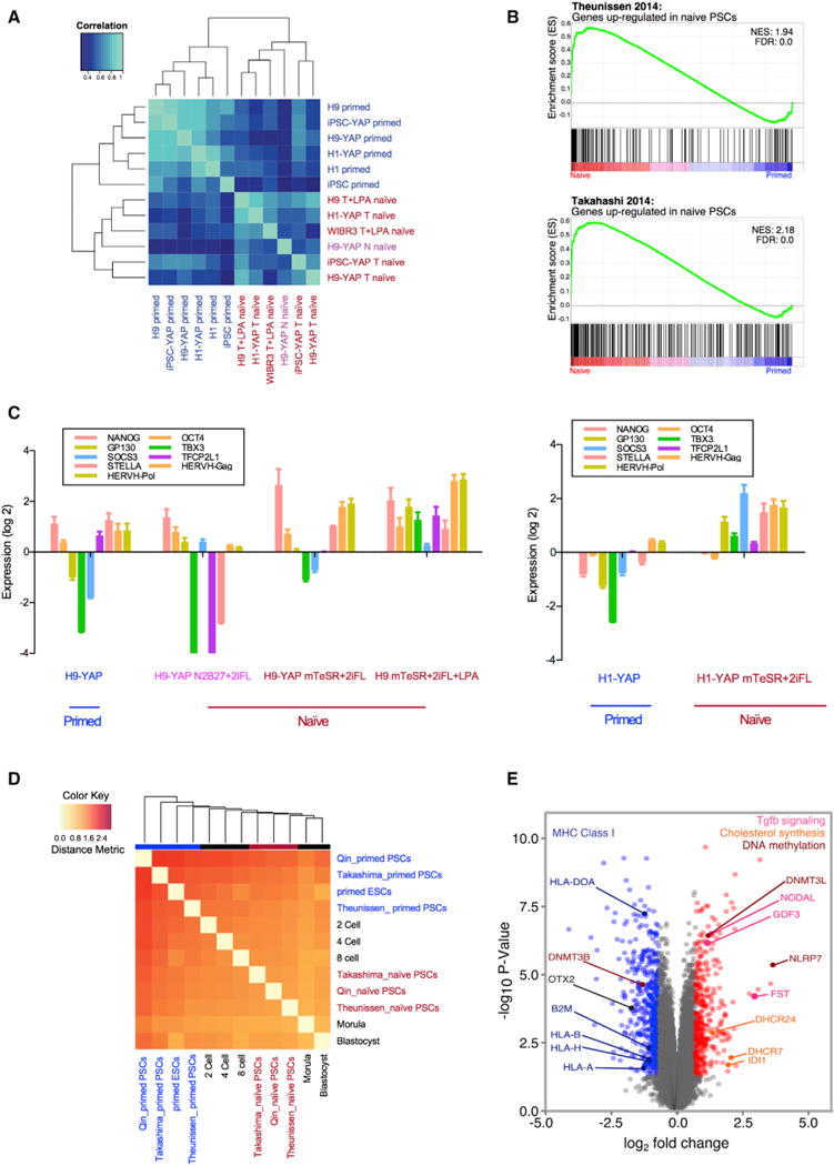
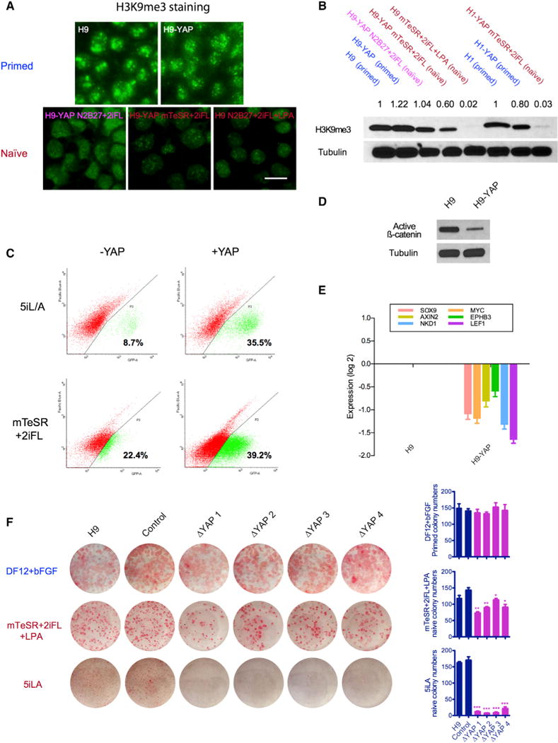
The human naive pluripotent stem cell (PSC) state, corresponding to a pre-implantation stage of development, has been difficult to capture and sustain in vitro. We report that the Hippo pathway effector YAP is nuclearly localized in the inner cell mass of human blastocysts. Overexpression of YAP in human embryonic stem cells (ESCs) and induced PSCs (iPSCs) promotes the generation of naive PSCs. Lysophosphatidic acid (LPA) can partially substitute for YAP to generate transgene-free human naive PSCs. YAP- or LPA-induced naive PSCs have a rapid clonal growth rate, a normal karyotype, the ability to form teratomas, transcriptional similarities to human pre-implantation embryos, reduced heterochromatin levels, and other hallmarks of the naive state. YAP/LPA act in part by suppressing differentiation-inducing effects of GSK3 inhibition. CRISPR/Cas9-generated YAP(-/-) cells have an impaired ability to form colonies in naive but not primed conditions. These results uncover an unexpected role for YAP in the human naive state, with implications for early human embryology.

Keywords: Hippo pathway; Yes-associated protein (YAP); embryonic stem cells (ESCs); induced pluripotent stem cells (iPSCs); lysophosphatidic acid (LPA); naive pluripotency; pluripotent stem cells (PSCs).


Figures

Figure 1
Figure 1. YAP Overexpression Promotes the Generation of Human Naive ESCs
(A) YAP protein is localized to the nucleus in both the trophectoderm and the ICM of human blastocysts, as shown by immunofluorescence. White, DAPI; green, OCT4 (ICM); yellow, CDX2 (trophectoderm); red, YAP. Representative image of n = 16 human blastocysts. Scale bar, 20 μm. (B) In naive medium N2B27+2iFL, human H9 ESCs with YAP overexpression (H9-YAP) maintain expression of the pluripotency marker AP, while control H9 cells differentiate and do not express AP. Scale bar, 500 μm. (C) H9-YAP cells in N2B27+2iFL have a naive-specific dome-like colony morphology and show strong positive immunostaining for pluripotency markers SSEA3, SSEA4, TRA-1-60, TRA-1-81, OCT4, and NANOG. Black scale bar, 500 mm; white scale bar, 150 μm. (D) Growth curves of H9 in DF12+bFGF primed medium, H9-YAP in DF12+bFGF primed medium, and H9-YAP in N2B27+2iFL naive medium after trypsinization to single cells. Only H9-YAP cells in naive medium have a high proliferate rate. Error bars represent SD. (E) H9-YAP cells in N2B27+2iFL have a normal female karyotype (46, XX), as evaluated after 20 passages in naive culture conditions. (F) H9-YAP cells in N2B27+2iFL are able to form teratomas comprising tissues derived from all three germ layers. (a) Adipocytes (mesoderm). (b) Neural tissue (ectoderm). (c) Epithelium (endoderm). White scale bar, 200 μm; black scale bar, 50 μm.
Figure 2
Figure 2. LPA Can Activate YAP and Promotes a Naive State of Pluripotency
(A) LPA increases the level of YAP in human H9 ESCs, as shown by immunofluorescence. Immunostaining was performed 2 days after adding LPA. Green, YAP; blue, DAPI. Scale bar, 50 μm. (B) Western blotting confirming that LPA increases YAP protein levels. Tubulin was used as loading control. (C) H9 cells in N2B27+2iFL+LPA naive medium have a naive-specific dome-like colony morphology and show strong positive immunostaining for pluripotency markers SSEA3, SSEA4, TRA-1-60, and TRA-1-81. Black scale bar, 500 μm; white scale bar, 150 μm. (D) H9-YAP cells in N2B27+2iFL are able to form teratomas comprising tissues derived from all three germ layers. (a) Gut-like epithelium (endoderm). (b) Cartilage (mesoderm). (c) Neural tissue (ectoderm). White scale bar, 200 μm; black scale bar, 50 μm.
Figure 3
Figure 3. Yin-PSCs and Lin-PSCs Have a Naive-like Transcriptional Profile Distinct from that of Primed PSCs
(A) Primed PSCs and Yin- and Lin-PSCs have distinct gene expression profiles, as shown by unsupervised hierarchical clustering. The top 1,000 genes with the highest coefficient of variation were used to cluster samples using Pearson correlation coefficients. Six human primed PSC samples include H9, H1, IMR-90 iPSCs, H9-YAP, H1-YAP, and IMR-90 iPSC-YAP cells. Six human naive PSC samples include H9-YAP in N2B27+2iFL (H9-YAP N naive), H9-YAP in mTeSR+2iFL (H9-YAP T naive), H1-YAP in mTeSR+2iFL (H1-YAP T naive), IMR-90 iPSC-YAP cells in mTeSR+2iFL (iPSC-YAP T naive), H9 in mTeSR+2iFL+LPA (H9 T+LPA naive), and WIBR3 in mTeSR+2iFL+LPA (WIBR3 T+LPA naive). Primed cells are indicated in blue. H9-YAP in N2B27+2iFL naive medium is in pink, and all other naive cells are in red. (B) GSEA reveals that Yin-PSCs and Lin-PSCs have gene expression profiles concordant with naive PSCs from other studies. The upper panel is the enrichment plot for the gene set of Theunissen et al. (2014) upregulated in naive by Log2 FC > 3.0. The lower panel is the enrichment plot for the gene set of Takahashi et al. (2014) upregulated in naive by Log2 FC > 2.0. Vertical black bars represent the position of genes upregulated in naive cells in the Takashima et al. (2014) or Theunissen et al. (2014) studies, distributed along the differential expression values for the entire transcriptome in this study, and ranked from upregulated in Yin-PSCs and Lin-PSCs (red, left) to upregulated in parental primed PSCs (blue, right). (C) Yin-PSCs and Lin-PSCs express markers specific for naive pluripotency, as confirmed by qRT-PCR. Values were normalized to GAPDH and UBB and then compared to H9 in primed medium (left panel) or H1 in primed medium (right panel). Data are averages of triplicate PCR reactions, and error bars represent SD. (D) Hierarchical clustering shows that naive Yin-PSCs and Lin-PSCs are similar to naive cells from two other studies (Takashima et al., 2014; Theunissen et al., 2014) and to human pre-implantation embryos. In vivo states at various developmental stages (Yan et al., 2013) are included in the clustering. (E) Volcano plot of significantly differentially expressed genes among the six human naive PSC samples and the six human primed PSC samples. Highlighted in red are genes with aLog2 FC > 0.7 in naive and p < 0.05. In blue are genes with a Log2 FC < −0.7 and p < 0.05 in naive. See text for a description of the specific genes indicated.
Figure 4
Figure 4. YAP Regulates the Human Naive State and Acts in Part by Modulating Wnt Signaling
(A) H3K9me3 is strongly reduced in naive H9 ESCs, as seen by immunofluorescence. Upper panel: H9 and H9-YAP in DF12+bFGF primed medium. Lower panel: H9-YAP in N2B27+2iFL naive medium, H9-YAP in mTeSR+2iFL naive medium, and H9 in mTeSR+2iFL+LPA naive medium. Scale bar, 20 μm. (B) Decreased total amount of H3K9me3 in Yin-PSCs and Lin-PSCs was confirmed by western blotting. Tubulin was used as loading control. Values indicate densitometry analysis of the H3K9me3 level normalized to tubulin. (C) Flow cytometric analysis of the proportion of OCT4-ΔPE-GFP+ WIBR3 cells with or without YAP overexpression in 5i/L/A (upper panels) or mTeSR+2iFL (lower panels) media. Cells were expanded in bulk and analyzed at passage 3, in the absence of colony picking. YAP overexpression increases the ratio of OCT4-ΔPE-GFP + cells in both media. (D) YAP overexpression decreases levels of unphosphorylated (active) β-catenin, as shown by western blotting. Tubulin was used as loading control. (E) YAP overexpression decreases the expression of Wnt target genes, as shown by qRT-PCR. Values were normalized to GAPDH and UBB and then compared to H9. Data are averages of triplicate PCR reactions, and error bars represent SD. (F) YAP knockout impairs the ability of ESCs to form naive colonies. H9 controls and four clones of CRISPR/Cas9-generated YAP−/− cells cultured in DF12+bFGF (primed), mTeSR+2iFL+LPA (naive), and 5i/L/A (naive, Theunissen et al., 2014) were trypsinized to single cells, counted, and plated onto MEFs in the presence of ROCK inhibitor. Seven days later, AP staining was performed and colony numbers were counted. Error bars represent SD. *p < 0.05; **p < 0.01; ***p < 0.001.

References

    1. Azzolin L, Panciera T, Soligo S, Enzo E, Bicciato S, Dupont S, Bresolin S, Frasson C, Basso G, Guzzardo V, et al. YAP/TAZ incorporation in the β-catenin destruction complex orchestrates the Wnt response. Cell. 2014;158:157–170. - PubMed
    1. Blair K, Wray J, Smith A. The liberation of embryonic stem cells. PLoS Genet. 2011;7:e100. 2019. - PMC - PubMed
    1. Buecker C, Chen HH, Polo JM, Daheron L, Bu L, Barakat TS, Okwieka P, Porter A, Gribnau J, Hochedlinger K, Geijsen N. A murine ESC-like state facilitates transgenesis and homologous recombination in human pluripotent stem cells. Cell Stem Cell. 2010;6:535–546. - PMC - PubMed
    1. Buecker C, Srinivasan R, Wu Z, Calo E, Acampora D, Faial T, Simeone A, Tan M, Swigut T, Wysocka J. Reorganization of enhancer patterns in transition from naive to primed pluripotency. Cell Stem Cell. 2014;14:838–853. - PMC - PubMed
    1. Bustamante-Marín X, Garness JA, Capel B. Testicular teratomas: an intersection of pluripotency, differentiation and cancer biology. Int J Dev Biol. 2013;57:201–210. - PubMed

Publication types

MeSH terms