. 2016 Mar 7:6:22741.
doi: 10.1038/srep22741.

Subcortical cytoskeleton periodicity throughout the nervous system

Affiliations

Subcortical cytoskeleton periodicity throughout the nervous system

Elisa D'Este et al. Sci Rep. .

Abstract

Superresolution fluorescence microscopy recently revealed a ~190 nm periodic cytoskeleton lattice consisting of actin, spectrin, and other proteins underneath the membrane of cultured hippocampal neurons. Whether the periodic cytoskeleton lattice is a structural feature of all neurons and how it is modified when axons are ensheathed by myelin forming glial cells is not known. Here, STED nanoscopy is used to demonstrate that this structure is a commonplace of virtually all neuron types in vitro. To check how the subcortical meshwork is modified during myelination, we studied sciatic nerve fibers from adult mice. Periodicity of both actin and spectrin was uncovered at the internodes, indicating no substantial differences between unmyelinated and myelinated axons. Remarkably, the actin/spectrin pattern was also detected in glial cells such as cultured oligodendrocyte precursor cells. Altogether our work shows that the periodic subcortical cytoskeletal meshwork is a fundamental characteristic of cells in the nervous system and is not a distinctive feature of neurons, as previously thought.


Conflict of interest statement

SWH owns shares of the companies Abberior and Abberior Instruments GmbH, producing superresolution STED dyes and microscopes, respectively.

Figures
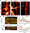
Figure 1
Figure 1. Subcortical cytoskeleton periodicity in the central nervous system.
(a–c) Representative live STED images of cortical neurons (a), striatal neurons (b) and cerebellar granule cells (c) stained with SiR-Actin and neurofascin (insets). (d) Schematic drawing of a retinal bipolar cell with a long axon and short dendrites. (e,g) Two-color STED images of phalloidin- and betaII spectrin- stained retinal bipolar cell axon (e) and dendrite (g), highlighting the cytoskeletal periodic organization. (f) Line profile along the dashed line in (e). (h) Line profile along the dashed line in (g). All image data was smoothed. All scale bars: 1 μm.
Figure 2
Figure 2. Subcortical cytoskeleton periodicity in the peripheral nervous system.
(a) Living DRG neurons stained with SiR-Actin and imaged with STED nanoscopy at 6 DIV show long-range actin periodicity. (a’) Close-up of the area indicated in (a). (b) BetaII spectrin shows short-range periodic order in DRG neurons at 6 DIV. (c) BetaII spectrin and phalloidin co-staining shows an alternating pattern (6 DIV). (d) Line profile of phalloidin and betaII spectrin intensities along the dashed line in (c). (e) Phalloidin staining of fixed DRG shows the presence of the actin pattern already at 2 DIV. (f) Sciatic nerve stained with phalloidin and imaged with STED shows actin periodic organization at the internode. (g) BetaII spectrin periodic organization along the axon at internodal region. (h) Line profile of phalloidin intensity along the dashed line in (f). (i) STED image of another phalloidin stained internode highlights actin presence along the axon (periodic) and on the outermost myelin layer. (j) AnkyrinG staining at a node of Ranvier of a thin sliced sciatic nerve. All image data was smoothed. All scale bars: 1 μm.
Figure 3
Figure 3. The subcortical cytoskeleton in glial cells.
(a,b) Representative STED images of phalloidin stained microglial cells at 3 DIV (a) and astrocytes at 1 DIV (b). (c) Living oligodendrocytes stained with SiR-Actin and imaged with STED nanoscopy at 5 DIV. (c’,c’’,c’’’) Close-ups of the areas indicated in (c). (d) BetaII spectrin and phalloidin co-staining in oligodendrocytes at 6 DIV. (d’) Close-up of the area indicated in (d) shows an alternating pattern of actin and betaII spectrin. (e) Line profile along the dashed line in (d’). All image data was smoothed. All scale bars: 1 μm.
Figure 4
Figure 4. Comparison of actin spacing in the different neuronal cell types.
Box plot of the actin inter-peak spacing in the different cells. All measurements have been performed on the axons (when applicable). HPN: hippocampal neurons. Data for HPN axons, HPN axon phalloidin, and HPN dendrites taken from the experiments performed in (npeaks = 65, ncells = 7 for HPN axons; npeaks = 80, ncells = 5 for HPN axons Phall; npeaks = 50, ncells = 4 for HPN Dendrites). Data for the other cell types and tissues are reported in the text.

References

    1. Schmitt H., Gozes I. & Littauer U. Z. Decrease in levels and rates of synthesis of tubulin and actin in developing rat brain. Brain Res 121, 327–342, (1977). - PubMed
    1. Goodman S. R. et al. A spectrin-like protein from mouse brain membranes: immunological and structural correlations with erythrocyte spectrin. Cell Motil 3, 635–647 (1983). - PubMed
    1. Bennett V., Davis J. & Fowler W. E. Brain spectrin, a membrane-associated protein related in structure and function to erythrocyte spectrin. Nature 299, 126–131 (1982). - PubMed
    1. Xu K., Zhong G. & Zhuang X. Actin, spectrin, and associated proteins form a periodic cytoskeletal structure in axons. Science 339, 452–456 (2013). - PMC - PubMed
    1. Zhong G. et al. Developmental mechanism of the periodic membrane skeleton in axons. Elife 3 (2014). - PMC - PubMed

Publication types

LinkOut - more resources