. 2016 Jul 1;23(7):773-91.
doi: 10.5551/jat.31401. Epub 2016 Mar 5.

Involvement of Angiotensin II Type 1 and 2 Receptors in Gelatinase Regulation in Human Carotid Atheroma in vitro

Affiliations

Involvement of Angiotensin II Type 1 and 2 Receptors in Gelatinase Regulation in Human Carotid Atheroma in vitro

Paula Clancy et al. J Atheroscler Thromb. .

Abstract

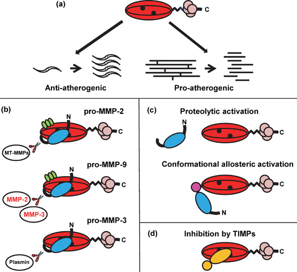
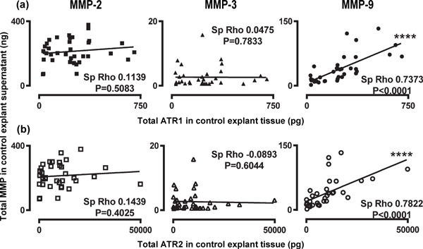
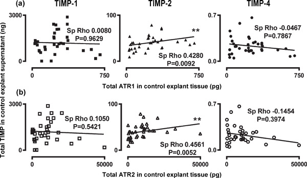
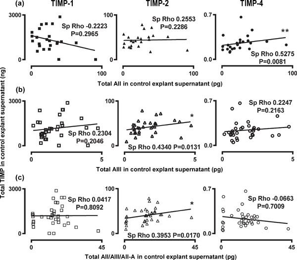
Aim: Matrix metalloproteinases (MMPs), angiotensin II (AII) and its receptors are implicated in atherosclerotic plaque instability, however the roles of the two receptor subtypes, ATR1 and ATR2, in MMP regulation remain uncertain. In this study, we investigated the effect of ATR1 and ATR2 blockade on the expression and activity of MMP-2, MMP-3 and MMP-9, in human carotid atheroma.

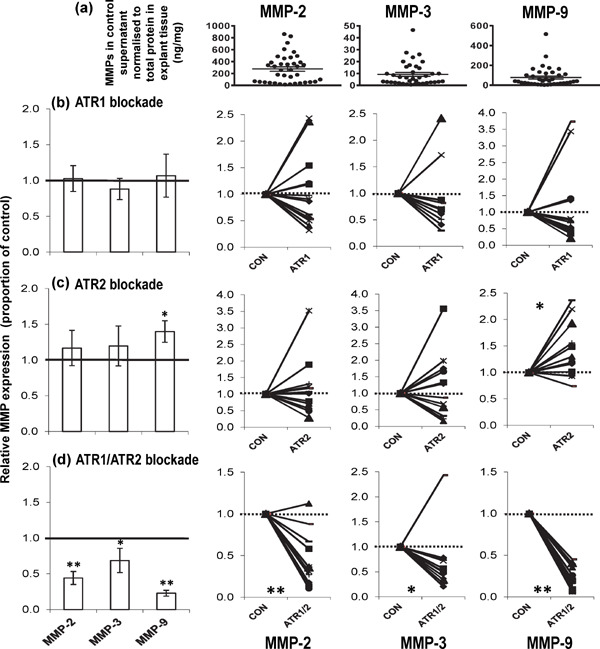
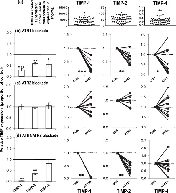
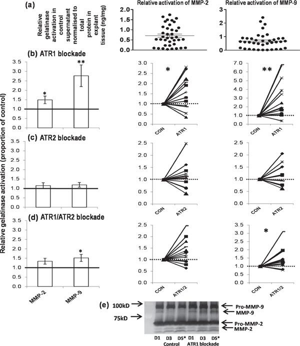
Methods: Atheroma samples (n=36) were obtained from patients undergoing carotid endarterectomy. The effects of ATR1 (irbesartan), ATR2 (PD123319) and combined ATR1 and ATR2 blockade on the expression and activity of the MMPs and the expression of tissue inhibitors of metalloproteinases (TIMPs) were investigated in explant culture experiments. Paired atheroma samples were incubated with the intervention or media control for 4 days. Protein levels (MMP-2, MMP-3, MMP-9, TIMP-1, TIMP-2, TIMP-4, ATR1 and ATR2) were determined by ELISA. Overall gelatinase activity and specific activation were measured by chromogenic activity assays and zymography, respectively.

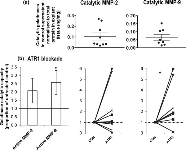
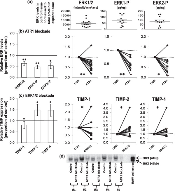
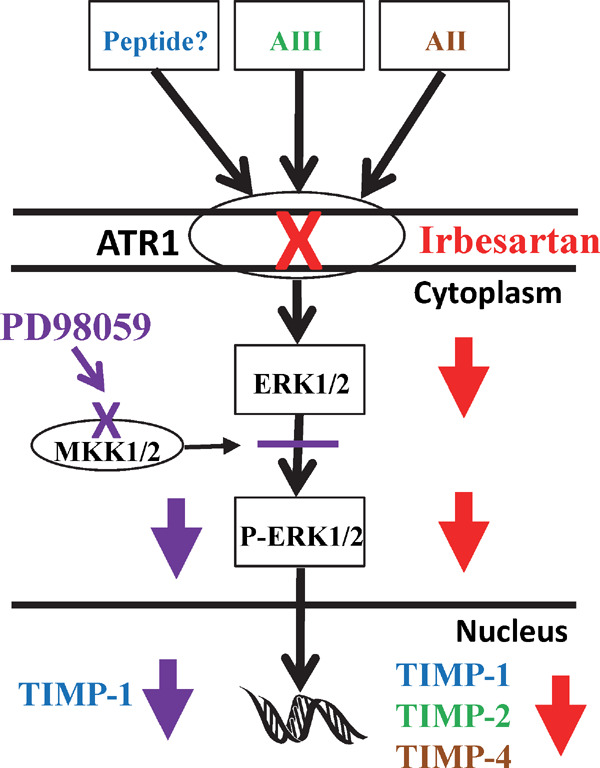
Results: ATR1 blockade, but not ATR2 blockade significantly reduced TIMP-1, TIMP-2 and TIMP-4 expression in atheroma supernatant. Combined ATR1 and ATR2 blockade significantly reduced MMP-2, MMP-3 and MMP-9 expression. MMP-2 and MMP-9 relative activation, and overall MMP-9 catalytic capacity were significantly increased by ATR1 blockade.

Conclusions: Our findings suggest that ATR1 blockade reduces TIMP expression and increases gelatinase activity in human carotid atheroma.


Figures

Fig. 1.
Fig. 1.
Structure and function of MMPs and role in atherosclerosis. (a) MMPs are potentially involved in SMC migration and proliferation (anti-atherogenic activity) and also collagen and elastin degradation (pro-atherogenic activity) within the atheroma. (b) Domain structure for the MMPs. The archetypal domain format is an N-terminal signal sequence+propeptide (blue) which blocks substrate access to the active site by forming a bond between the Zn2+ ions (black) in the catalytic site and Cys 73 in the propeptide (cysteine “switch”) + catalytic domain (red) + hinged region + 4 hemopexin domains (pink) + tail sequence. MMP-3 has this domain format, whereas MMP-2 and MMP-9 have 3 extra fibronectin type II regions within the catalytic domain (green). Key enzymes that can activate the MMPs are also shown including family members investigated in this study (red text). (c) MMPs are heavily regulated with activation occurring when the regulating Cys-Zn2+ bond is broken exposing the catalytic site. This can be by protease degradation or allosteric means. (d) Inhibition occurs when TIMPs bind to the active site. All the TIMPs are capable of binding to all the MMPs. The overall catalytic capacity of the MMPs is a finely tuned balance between all these factors.
Fig. 2.
Fig. 2.
Changes in MMP expression following angiotensin receptor blockade. Mean ± SEM of (a) MMP-2, MMP-3 and MMP-9 detected in the control culture supernatant, normalised to the amount of protein present in the explant tissue (n = 38, control samples from the 3 interventions). Relative expression of MMPs in human carotid atheroma supernatant after 4d culture with and without (b) ATR1 (irbesartan, 2.3 mmol/L, n = 14 pairs); (c) ATR2 (PD123319, 1 µmol/L, n = 12 pairs); and (d) ATR1/ATR2 (irbesartan, 2.3 mmol/L and PD123319, 1 µmol/L, n = 12 pairs) blockade. Shown are the mean values ± SEM of the ratio of enzyme (normalised to total protein) secreted by paired atheroma samples incubated with intervention relative to control. Also shown are the individual matched pairs. Total MMP expression was determined using ELISA. **P < 0.01; *P < 0.05 using Wilcoxon's paired test. (1 = no change, > 1 increased, < 1 decreased with intervention)
Fig. 3.
Fig. 3.
Changes in TIMP expression following angiotensin receptor blockade. Mean ± SEM of (a) TIMP-1, TIMP-2 and TIMP-4 detected in the control culture supernatant, normalised to the amount of protein present in the explant tissue (n = 50, control samples from the 4 interventions). Relative expression of TIMPs in human carotid atheroma supernatant after 4d culture with and without (b) ATR1 (irbesartan, 2.3 mmol/L, n = 14 pairs); (c) ATR2 (PD123319, 1 µmol/L, n = 12 pairs); and (d) ATR1/ATR2 (irbesartan, 2.3 mmol/L and PD123319, 1 µmol/L, n = 12 pairs) blockade. Shown are the mean values ± SEM of the ratio of enzyme (normalised to total protein) secreted by paired atheroma samples incubated with intervention relative to control. Also shown are the individual matched pairs. TIMP expression was determined using ELISA. ***P < 0.001; **P < 0.01; *P < 0.05 using Wilcoxon's paired test. (1 = no change, > 1 increased, < 1 decreased with intervention)
Fig. 4.
Fig. 4.
Changes in relative gelatinase activation following angiotensin receptor blockade. Mean ± SEM of (a) relative activated MMP-2 and relative activated MMP-9 detected in the control culture supernatant, normalised to the amount of protein present in the explant tissue (n = 36, control samples from the 3 interventions). Relative activation of MMP-2 and -9 in human carotid atheroma supernatant after 4d culture with and without (b) ATR1 (irbesartan, 2.3 mmol/L, n = 14 pairs), (c) ATR2 (PD123319, 1 µmol/L, n = 12 pairs) and (d) ATR1/ATR2 (irbesartan, 2.3 mmol/L and PD123319, 1 µmol/L, n = 12 pairs) blockade. Shown are the mean values ± SEM of the ratio of cleaved enzyme (Fig. 1c) to pro-enzyme (Fig. 1b) (normalised to total protein) secreted by paired atheroma samples incubated with intervention relative to control. Also shown are the individual matched pairs. (e) Relative levels of gelatinase activation were determined using zymography. The asterisked day 5 samples (D5*) were the ones used in the analyses. NB the day 1 samples (D1) are actually the same for both control and ATR1 blockade as there is an initial 24h “media only” pre-culture step to reduce the impact of any medications the patient may have been taking before implementation of the interventions in culture. This gel also demonstrates that gelatinase expression does not decline in the 5 days of culture as all the control samples are consistent for all three time points. **P < 0.01; *P < 0.05 using Wilcoxon's paired test. (1 = no change, > 1 increased, < 1 decreased with intervention)
Fig. 5.
Fig. 5.
Changes in overall gelatinase catalytic capacity following ATR1 blockade. Mean ± SEM of (a) catalytically active MMP-2 (n = 9) and catalytically active MMP-9 (n = 8) (active MMP-2 and -9 were not detectable in all sample pairs) detected in the control culture supernatant, normalised to the amount of protein present in the explant tissue. Overall catalytic capacity of MMP-2 and -9 in human carotid atheroma supernatant after 4d culture with and without ATR1 blockade (irbesartan, 2.3 mmol/L, n = 9 and 8 pairs, respectively). Shown are the mean values ± SEM of the ratio of active enzyme (normalised to total protein) secreted by paired atheroma samples incubated with intervention relative to control. Also shown are the individual matched pairs. Overall catalytic capacity for each gelatinase was determined by a chromogenic assay, which only detects proteolytically cleaved and activated MMPs that are not complexed to a TIMP (Fig. 1c). *P < 0.05 using Wilcoxon's paired test. (1 = no change, > 1 increased with intervention).
Fig. 6.
Fig. 6.
MMP-9 up-regulation in the serum stimulated, mixed, healthy primary vascular cell model. (a) Expression of MMP-9 in the culture supernatant after 4d of culture using various combinations of healthy EC, SMC, BCs and serum. Shown are the mean values from triplicate cultures ± SEM. Expression of MMP-9 in the mixed, healthy primary vascular cell culture (EC, SMC, BC and serum) supernatant after 4d of culture with (b) titrated ATR1 blockade, (b) combined ATR1/ATR2 blockade, and (c) titrated ATR2 blockade. Shown are the mean values from triplicate cultures ± SEM.
Fig. 7.
Fig. 7.
Changes in ERK and TIMP expression following various interventions. Mean ± SEM of (a) ERK1/2, ERK1-P and ERK2-P detected in the control culture tissue, normalised to the amount of total protein present in the explant tissue (n = 14 control samples from the ATR1 intervention). Relative expression of (b) ERKs in human carotid atheroma tissue after 4d culture with and without ATR1 (irbesartan, 2.3 mmol/L, n = 14 pairs) blockade and (c) TIMPs in the human carotid atheroma culture supernatant after 4d culture with and without ERK1/2 activity (PD98059, 20 µmol/L, n = 12 pairs) blockade. Shown are the mean values ± SEM of the ratio of enzyme (normalised to total protein) secreted by paired atheroma samples incubated with intervention relative to control. Also shown are the individual matched pairs. TIMP and activated ERK levels were determined using ELISA. (d) ERK1/2 expression was determined using Western blotting (load 30 µg total protein in 10 µL loading buffer). **P < 0.01; *P < 0.05 using Wilcoxon's paired test. (1 = no change, > 1 increased, < 1 decreased with intervention)
Fig. 8.
Fig. 8.
Model of molecular mechanism of TIMP expression with ATR1 blockade. Presented is a simplified pathway by which ATR1 blockade potentially leads to reduced TIMP production. ATR1 has a number of angiotensin peptide agonists that are blocked by nonpeptide diphenylimidazole antagonists, such as irbesartan (red cross). The correlation studies suggest that TIMP-2 expression is dependent on AIII binding to ATR1 and TIMP-4 expression is dependent on AII binding to ATR1. Blockade of ATR1 led to a decrease in expression of the TIMPs and a concomitant decrease in the expression of ERK1/2 in the tissue. Activation of ERK1/2 was blocked by PD98059, which binds to MKK1/2, inactivating the enzymes capacity to phosphorylate ERK1/2 (purple cross). Blockade of ERK1/2 activity also led to a decrease in TIMP-1 expression suggesting that TIMP-1 expression is down-regulated with ATR1 blockade via a decrease in ERK1/2 expression.
Supplemental Fig. 1.
Correlation of MMPs in control explant culture supernatant with ATRs in the control explant tissue. Scatter graphs for Spearman's rho correlations between MMPs and (a) ATR1 and (b) ATR2.
Supplemental Fig. 2.
Correlation of TIMPs in control explant culture supernatant with ATRs in the control explant tissue. Scatter graphs for Spearman's rho correlations between TIMPs and (a) ATR1 and (b) ATR2.
Supplemental Fig. 3.
Correlation of TIMPs and angiotensin peptides in control explant culture supernatant. Scatter graphs for Spearman's rho correlations between TIMPs and (a) AII, (b) AIII and (c) AII/AIII/AII-A.
Supplemental Fig. 4.
Correlation of TIMPs in matched control and ATR1 blockade sample supernatants with ERK1/2 signalling proteins in the explant tissue. Scatter graphs for Spearman's rho correlations between TIMPs and ERK1/2 signalling proteins.

Similar articles

Cited by

References

    1. Golledge J, Greenhalgh RM, Davies AH: The symptomatic carotid plaque. Stroke, 2000; 31: 774-781 - PubMed
    1. Dollery CM, Libby P: Atherosclerosis and proteinase activation. Cardiovasc Res, 2005; 69: 625-635 - PubMed
    1. Sukhova GK, Schonbeck U, Rabkin E, Schoen FJ, Poole AR, Billinghurst RC, Libby P: Evidence for increased Collagenolysis by interstitial Collagenases-1 and -3 in Vulnerable Human Atheromatous Plaques. Circulation, 1999; 99: 2503-2509 - PubMed
    1. Shah PK: Mechanisms of plaque vulnerability and rupture. J Am Coll Cardiol, 2003; 41: 15S-22S - PubMed
    1. Pardo A, Selman M: MMP-1: the elder of the family. Int J Biochem Cell Biol, 2005; 37: 283-288 - PubMed

MeSH terms