. 2016 May;25(5):1069-74.
doi: 10.1002/pro.2916. Epub 2016 Mar 24.

Structural characterization of NRAS isoform 5

Affiliations

Structural characterization of NRAS isoform 5

Joseph Markowitz et al. Protein Sci. 2016 May.

Abstract

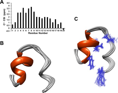
It was recently discovered that the NRAS isoform 5 (20 amino acids) is expressed in melanoma and results in a more aggressive cell phenotype. This novel isoform is responsible for increased phosphorylation of downstream targets such as AKT, MEK, and ERK as well as increased cellular proliferation. This structure report describes the NMR solution structure of NRAS isoform 5 to be used as a starting point to understand its biophysical interactions. The isoform is highly flexible in aqueous solution, but forms a helix-turn-coil structure in the presence of trifluoroethanol as determined by NMR and CD spectroscopy.

Keywords: NMR; NRAS; isoform; melanoma.


Figures

Figure 1
Figure 1
NRAS isoform 5 is a flexible peptide that is induced to form an α helix in the presence of trifluoroethanol. A) The CD dichroism spectrum of NRAS isoform 5 in the absence of TFE is presented and represents a spectrum that would be expected for a random coil. B) The CD dichroism spectrum of NRAS isoform 5 in the presence of 56% TFE is presented and represents a spectrum with α helical components. In the Supporting Information data, the complete TFE titration is presented.
Figure 2
Figure 2
NRAS isoform 5 main structural features include a helix, turn, and coil. A) Cα chemical shift Index (CSI) is presented. This CSI is higher between residues 4 and 9 compared with other regions. Together with NOE data it was determined that the alpha helix extends from Tyr 4 to Gly 10. B) Twenty‐five independently derived NMR structures from the program CYANA are superimposed. C) The structural ensemble is shown with the side chains of residues (Tyr 4, Val 8, Val 9, Val 14, and Trp 20) important for the tertiary fold of NRAS isoform 5.

References

    1. Linos E, Swetter SM, Cockburn MG, Colditz GA, Clarke CA (2009) Increasing burden of melanoma in the United States. J Invest Dermatol 129:666–1674. - PMC - PubMed
    1. Siegel RL, Miller KD, Jemal A (2015) Cancer Stat 65:5–29. - PubMed
    1. Flaherty KT, Infante JR, Daud A, Gonzalez R, Kefford RF, Sosman J, Hamid O, Schuchter L, Cebon J, Ibrahim N, Kudchadkar R, III Burris HA, , Falchook G, Algazi A, Lewis K, Long GV, Puzanov I, Lebowitz P, Singh A, Little S, Sun P, Allred A, Ouellet D, Kim KB, Patel K Weber J (2012) Combined BRAF and MEK inhibition in melanoma with BRAF V600 mutations. New Engl J Med 367:1694–1703. - PMC - PubMed
    1. Hartsough EJ, Basile KJ, Aplin AE (2014) Beneficial effects of RAF inhibitor in mutant BRAF splice variant‐expressing melanoma. Mol Cancer Res 12:795–802. - PMC - PubMed
    1. Basile KJ, Le K, Hartsough EJ, Aplin AE (2014) Inhibition of mutant BRAF splice variant signaling by next‐generation, selective RAF inhibitors. Pigment Cell Melanoma Res 27:479–484. - PMC - PubMed

Publication types

LinkOut - more resources