. 2016 Jun;14(6):1238-48.
doi: 10.1111/jth.13307. Epub 2016 Apr 5.

Tissue factor deficiency increases alveolar hemorrhage and death in influenza A virus-infected mice

Affiliations

Tissue factor deficiency increases alveolar hemorrhage and death in influenza A virus-infected mice

S Antoniak et al. J Thromb Haemost. 2016 Jun.

Erratum in

  • Corrigendum.
    [No authors listed] [No authors listed] J Thromb Haemost. 2016 Dec;14(12):2565. doi: 10.1111/jth.13521. Epub 2016 Dec 1. J Thromb Haemost. 2016. PMID: 28026156 No abstract available.

Abstract

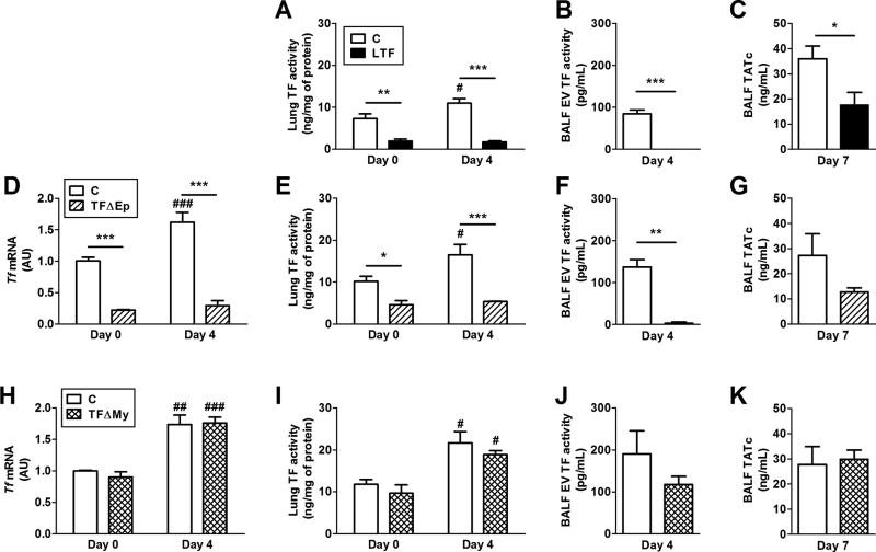
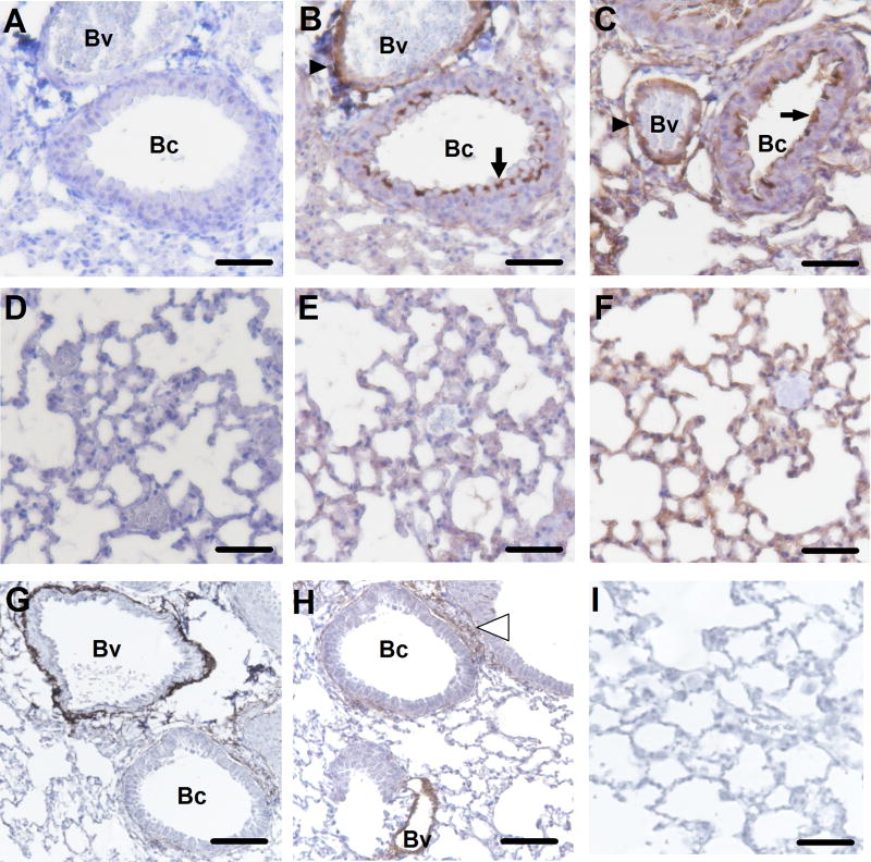
Essentials H1N1 Influenza A virus (IAV) infection is a hemostatic challenge for the lung. Tissue factor (TF) on lung epithelial cells maintains lung hemostasis after IAV infection. Reduced TF-dependent activation of coagulation leads to alveolar hemorrhage. Anticoagulation might increase the risk for hemorrhages into the lung during severe IAV infection.

Summary: Background Influenza A virus (IAV) infection is a common respiratory tract infection that causes considerable morbidity and mortality worldwide. Objective To investigate the effect of genetic deficiency of tissue factor (TF) in a mouse model of IAV infection. Methods Wild-type mice, low-TF (LTF) mice and mice with the TF gene deleted in different cell types were infected with a mouse-adapted A/Puerto Rico/8/34 H1N1 strain of IAV. TF expression was measured in the lungs, and bronchoalveolar lavage fluid (BALF) was collected to measure extracellular vesicle TF, activation of coagulation, alveolar hemorrhage, and inflammation. Results IAV infection of wild-type mice increased lung TF expression, activation of coagulation and inflammation in BALF, but also led to alveolar hemorrhage. LTF mice and mice with selective deficiency of TF in lung epithelial cells had low basal levels of TF and failed to increase TF expression after infection; these two strains of mice had more alveolar hemorrhage and death than controls. In contrast, deletion of TF in either myeloid cells or endothelial cells and hematopoietic cells did not increase alveolar hemorrhage or death after IAV infection. These results indicate that TF expression in the lung, particularly in epithelial cells, is required to maintain alveolar hemostasis after IAV infection. Conclusion Our study indicates that TF-dependent activation of coagulation is required to limit alveolar hemorrhage and death after IAV infection.

Keywords: hemorrhage; hemostasis; influenza A virus; pneumonia; tissue factor.


Conflict of interest statement

Conflict-of-interest disclosure: The authors declare no competing financial interests.

Figures

Figure 1
Figure 1. Influenza A infection increases TF expression and inflammation in the mouse lung
Wild-type mice were infected with influenza A virus (IAV) and samples were collected before and 1, 3, 4, 7 and 14 days after infection. (A) IAV genome levels in the lungs were quantified by real-time PCR and normalized to Rpl4 mRNA levels. Data are shown relative to day 3. (B) TF activity levels in the lung were measured using a one-stage clotting assay. (C) TF activity levels of extracellular vesicles (EV) isolated from the bronchoalveolar lavage fluid (BALF) were measured using a two-stage FXa generation assay. (D) Levels of thrombin-antithrombin complexes (TATc) in the BALF. (E) Levels of hemoglobin (Hb) in the BALF. (F) Levels of white blood cells (WBCs) in the BALF after IAV infection. (G) Levels of IL-1β in the BALF after IAV infection. (H) Levels of Ccl2 in the BALF after IAV infection. Data (mean ± SEM; n = 3–13 per group) were analyzed by 1-way ANOVA. Statistical significances are shown as *P<0.05, **P<0.01 and ***P<0.001 versus day 0.
Figure 2
Figure 2. Levels of TF expression in TF deficient mice before and after influenza A infection
(A–C) Low TF (LTF) mice, (D–G)TFfl/fl,SPC-Cre (TFΔEp) and (H–K) TFfl/fl,LysM-Cre (TFΔMy) mice and their respective control mice (referred as C) were infected with influenza A virus (IAV) and the lungs and bronchoalveolar lavage fluid (BALF) were collected. Levels of lung Tf mRNA (D and H) and TF activity (A, E and I), TF activity of BALF extracellular vesicles (EV) (B, F and J), and BALF thrombin-antithrombin complexes (TATc) (C, G and K) are shown. Controls are shown in white bars and experimental mice are shown in black (LTF), hatched (TFΔEp), or cross-hatched (TFΔMy) bars. Levels of Tf mRNA in the lungs were quantified by real-time PCR before and 4 days after IAV infection. Data were normalized to Rpl4 mRNA levels. Levels of the uninfected controls were set to 1. Data (mean ± SEM; n = 3–7 for day 0, n =3–10 for day 4, and n = 3–8 for day 7) were analyzed by 2-way ANOVA or Student’s t-test. Statistical significance is shown as *P<0.05, **P<0.01 and ***P<0.001 between groups or #P<0.05, ##P<0.01 and ###P<0.001 versus uninfected control of the respective genotypes.
Figure 3
Figure 3. Influenza A infection increases TF protein expression in the lung
We analyzed TF antigen expression in the lungs of uninfected (B, E) and infected (7 dpi) wild-type mice (C, F), and infected TFΔEp mice (G–I) by immunohistochemistry. Tissue sections were incubated with (B, C, E, F–I) or without (A and D) a goat anti-mouse TF polyclonal antibody. The black arrows indicate TF expression in the epithelium of bronchi (Bc). The arrowheads indicate TF staining in adventitial cells of blood vessels (Bv). The white arrow indicates TF expression in adventitial cells surrounding a bronchiole. Original magnification x200. Scale bar is 100 µm.
Figure 4
Figure 4. Effect of genetic deficiency of TF on alveolar hemorrhage in mice after influenza A infection
(A) Low TF (LTF) mice, (B) Tffl/fl,SPC-Cre (TFΔEp), (C) Tffl/fl,LysM-Cre (TFΔMy) and (D) Tffl/fl,Tie2-Cre (TFΔEc) mice and their respective control mice (referred as C) were infected with influenza A virus (IAV), and the bronchoalveolar lavage fluid (BALF) was collected before and 7 days after infection. Gross appearance of BALF and levels of hemoglobin (Hb) in BALF before and after IAV infection are shown. Controls are shown as white bars and experimental mice are shown as black bars (LTF), hatched (TFΔEp), cross-hatched (TFΔMy), or vertical-striped (TFΔEc) bars. Data (mean ± SEM; 3– 11) were analyzed by Student’s t-test. Statistical significance is shown as *P<0.05 between groups.
Figure 5
Figure 5. Effect of a global or cell type-specific deficiency of TF on mortality of mice after influenza A infection
(A and B) Low TF (LTF), (C and D) Tffl/fl,SPC-Cre (TFΔEp), (E and F), Tffl/fl,LysM-Cre (TFΔMy) and (G and H) Tffl/fl,Tie2-Cre (TFΔEc) mice and their respective control mice (referred as C) were infected with influenza A virus (IAV) and observed for 14 days. Survival rates (A, C, E and G) and changes in body weights of infected mice (B, D, F and H) are shown. Body weights before infection were set to 100% and did not differ significantly between genotypes. Data (mean ± SEM; n = 9–10 for A and B, n = 17–30 for C and D, n = 7–8 for E and F, and n = 14–24 for G and H) were analyzed by log-rank test (A, C, E and G) or by 2-way ANOVA (B, D, F and H). Statistical significance is shown as *P<0.05 and **P<0.01 between groups.

References

    1. Kroger AT, Atkinson WL, Marcuse EK, Pickering LK Advisory Committee on Immunization Practices Centers for Disease C, Prevention. General recommendations on immunization: recommendations of the Advisory Committee on Immunization Practices (ACIP) MMWR Recomm Rep. 2006;55:1–48. - PubMed
    1. Antoniak S, Mackman N. Multiple roles of the coagulation protease cascade during virus infection. Blood. 2014;123:2605–13. - PMC - PubMed
    1. Geisbert TW, Young HA, Jahrling PB, Davis KJ, Kagan E, Hensley LE. Mechanisms underlying coagulation abnormalities in ebola hemorrhagic fever: overexpression of tissue factor in primate monocytes/macrophages is a key event. J Infect Dis. 2003;188:1618–29. - PubMed
    1. Funderburg NT, Mayne E, Sieg SF, Asaad R, Jiang W, Kalinowska M, Luciano AA, Stevens W, Rodriguez B, Brenchley JM, Douek DC, Lederman MM. Increased tissue factor expression on circulating monocytes in chronic HIV infection: relationship to in vivo coagulation and immune activation. Blood. 2010;115:161–7. - PMC - PubMed
    1. Huerta-Zepeda A, Cabello-Gutierrez C, Cime-Castillo J, Monroy-Martinez V, Manjarrez-Zavala ME, Gutierrez-Rodriguez M, Izaguirre R, Ruiz-Ordaz BH. Crosstalk between coagulation and inflammation during Dengue virus infection. Thromb Haemost. 2008;99:936–43. - PubMed

Publication types

MeSH terms