Pretreatment with low-dose gadolinium chloride attenuates myocardial ischemia/reperfusion injury in rats
- PMID: 26948086
- PMCID: PMC4820799
- DOI: 10.1038/aps.2015.156
Pretreatment with low-dose gadolinium chloride attenuates myocardial ischemia/reperfusion injury in rats
Abstract
Aim: We have shown that low-dose gadolinium chloride (GdCl3) abolishes arachidonic acid (AA)-induced increase of cytoplasmic Ca(2+), which is known to play a crucial role in myocardial ischemia/reperfusion (I/R) injury. The present study sought to determine whether low-dose GdCl3 pretreatment protected rat myocardium against I/R injury in vitro and in vivo.
Methods: Cultured neonatal rat ventricular myocytes (NRVMs) were treated with GdCl3 or nifedipine, followed by exposure to anoxia/reoxygenation (A/R). Cell apoptosis was detected; the levels of related signaling molecules were assessed. SD rats were intravenously injected with GdCl3 or nifedipine. Thirty min after the administration the rats were subjected to LAD coronary artery ligation followed by reperfusion. Infarction size, the release of serum myocardial injury markers and AA were measured; cell apoptosis and related molecules were assessed.
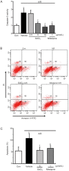
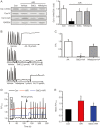
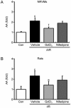
Results: In A/R-treated NRVMs, pretreatment with GdCl3 (2.5, 5, 10 μmol/L) dose-dependently inhibited caspase-3 activation, death receptor-related molecules DR5/Fas/FADD/caspase-8 expression, cytochrome c release, AA release and sustained cytoplasmic Ca(2+) increases induced by exogenous AA. In I/R-treated rats, pre-administration of GdCl3 (10 mg/kg) significantly reduced the infarct size, and the serum levels of CK-MB, cardiac troponin-I, LDH and AA. Pre-administration of GdCl3 also significantly decreased the number of apoptotic cells, caspase-3 activity, death receptor-related molecules (DR5/Fas/FADD) expression and cytochrome c release in heart tissues. The positive control drug nifedipine produced comparable cardioprotective effects in vitro and in vivo.
Conclusion: Pretreatment with low-dose GdCl3 significantly attenuates I/R-induced myocardial apoptosis in rats by suppressing activation of both death receptor and mitochondria-mediated pathways.
Figures


Similar articles
-
Protective effect of low dose gadolinium chloride against isoproterenol-induced myocardial injury in rat.Apoptosis. 2015 Sep;20(9):1164-75. doi: 10.1007/s10495-015-1147-8. Apoptosis. 2015. PMID: 26089194
-
Febuxostat pretreatment attenuates myocardial ischemia/reperfusion injury via mitochondrial apoptosis.J Transl Med. 2015 Jul 2;13:209. doi: 10.1186/s12967-015-0578-x. J Transl Med. 2015. PMID: 26136232 Free PMC article.
-
Astragaloside IV attenuates myocardial ischemia/reperfusion injury in rats via inhibition of calcium-sensing receptor-mediated apoptotic signaling pathways.Acta Pharmacol Sin. 2019 May;40(5):599-607. doi: 10.1038/s41401-018-0082-y. Epub 2018 Jul 20. Acta Pharmacol Sin. 2019. PMID: 30030530 Free PMC article.
-
Current Updates on Potential Role of Flavonoids in Hypoxia/Reoxygenation Cardiac Injury Model.Cardiovasc Toxicol. 2021 Aug;21(8):605-618. doi: 10.1007/s12012-021-09666-x. Epub 2021 Jun 10. Cardiovasc Toxicol. 2021. PMID: 34114196 Review.
-
Role of cell death in the progression of heart failure.Heart Fail Rev. 2016 Mar;21(2):157-67. doi: 10.1007/s10741-016-9532-0. Heart Fail Rev. 2016. PMID: 26872675 Review.
Cited by
-
Gadolinium and ruthenium red attenuate remote hind limb preconditioning-induced cardioprotection: possible role of TRP and especially TRPV channels.Naunyn Schmiedebergs Arch Pharmacol. 2016 Aug;389(8):887-96. doi: 10.1007/s00210-016-1251-5. Epub 2016 Apr 27. Naunyn Schmiedebergs Arch Pharmacol. 2016. PMID: 27118661
-
Novel Insight into the Role of Endoplasmic Reticulum Stress in the Pathogenesis of Myocardial Ischemia-Reperfusion Injury.Oxid Med Cell Longev. 2021 Mar 26;2021:5529810. doi: 10.1155/2021/5529810. eCollection 2021. Oxid Med Cell Longev. 2021. PMID: 33854692 Free PMC article. Review.
-
Gadolinium chloride pre-treatment reduces the inflammatory response and preserves intestinal barrier function in a rat model of sepsis.Exp Ther Med. 2021 Oct;22(4):1143. doi: 10.3892/etm.2021.10577. Epub 2021 Aug 9. Exp Ther Med. 2021. PMID: 34504589 Free PMC article.
-
MnO nanoparticles with potential application in magnetic resonance imaging and drug delivery for myocardial infarction.Int J Nanomedicine. 2018 Oct 8;13:6177-6188. doi: 10.2147/IJN.S176404. eCollection 2018. Int J Nanomedicine. 2018. PMID: 30323598 Free PMC article.
-
Liraglutide directly protects cardiomyocytes against reperfusion injury possibly via modulation of intracellular calcium homeostasis.J Geriatr Cardiol. 2017 Jan;14(1):57-66. doi: 10.11909/j.issn.1671-5411.2017.01.008. J Geriatr Cardiol. 2017. PMID: 28270843 Free PMC article.
References
-
- Frangogiannis NG, Smith CW, Entman ML. The inflammatory response in myocardial infarction. Cardiovasc Res 2002; 53: 31–47. - PubMed
-
- Sutton MG, Sharpe N. Left ventricular remodeling after myocardial infarction: pathophysiology and therapy. Circulation 2000; 101: 2981–8. - PubMed
-
- Mersmann J, Latsch K, Habeck K, Zacharowski K. Measure for ameasure-determination of infarct size in murine models of myocardial ischemia and reperfusion: a systematic review. Shock 2011; 35: 449–55. - PubMed
-
- Yellon DM, Hausenloy DJ. Myocardial reperfusion injury. N Engl J Med 2007; 357: 1121–35. - PubMed
Publication types
MeSH terms
Substances
LinkOut - more resources
Full Text Sources
Other Literature Sources
Research Materials
Miscellaneous

