

Vascular closure devices for femoral arterial puncture site haemostasis
- PMID: 26948236
- PMCID: PMC10372718
- DOI: 10.1002/14651858.CD009541.pub2
Vascular closure devices for femoral arterial puncture site haemostasis
Abstract
Background: Vascular closure devices (VCDs) are widely used to achieve haemostasis after procedures requiring percutaneous common femoral artery (CFA) puncture. There is no consensus regarding the benefits of VCDs, including potential reduction in procedure time, length of hospital stay or time to patient ambulation. No robust evidence exists that VCDs reduce the incidence of puncture site complications compared with haemostasis achieved through extrinsic (manual or mechanical) compression.
Objectives: To determine the efficacy and safety of VCDs versus traditional methods of extrinsic compression in achieving haemostasis after retrograde and antegrade percutaneous arterial puncture of the CFA.
Search methods: The Cochrane Vascular Trials Search Co-ordinator searched the Specialised Register (April 2015) and the Cochrane Central Register of Controlled Trials (CENTRAL) (2015, Issue 3). Clinical trials databases were searched for details of ongoing or unpublished studies. References of articles retrieved by electronic searches were searched for additional citations.
Selection criteria: We included randomised and quasi-randomised controlled trials in which people undergoing a diagnostic or interventional procedure via percutaneous CFA puncture were randomised to one type of VCD versus extrinsic compression or another type of VCD.
Data collection and analysis: Two authors independently extracted data and assessed the methodological quality of trials. We resolved disagreements by discussion with the third author. We performed meta-analyses when heterogeneity (I(2)) was < 90%. The primary efficacy outcomes were time to haemostasis and time to mobilisation (mean difference (MD) and 95% confidence interval (CI)). The primary safety outcome was a major adverse event (mortality and vascular injury requiring repair) (odds ratio (OR) and 95% CI). Secondary outcomes included adverse events.
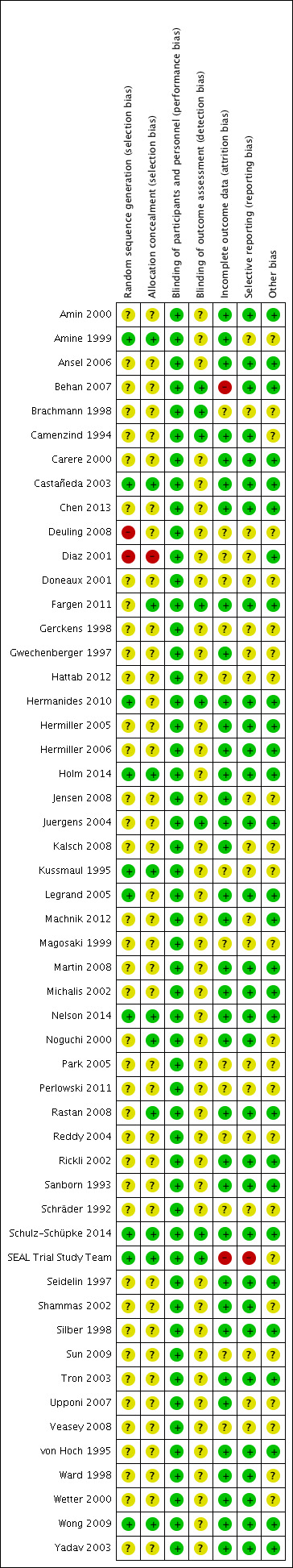
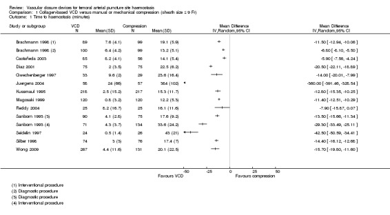
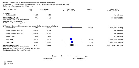
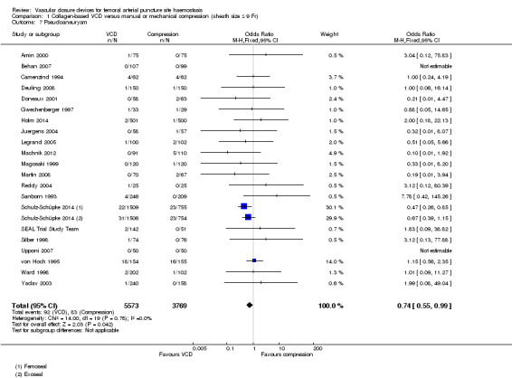
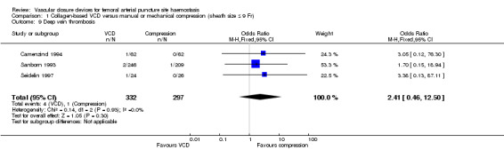
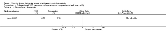
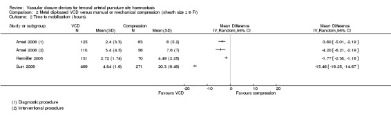
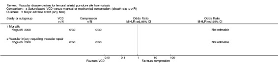
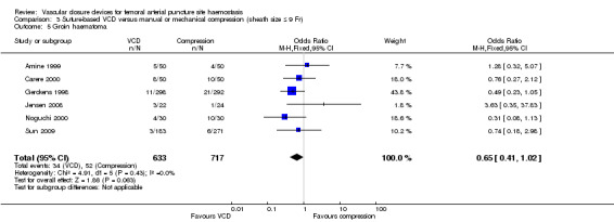
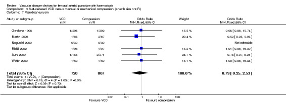
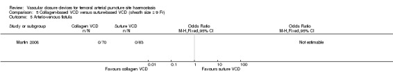
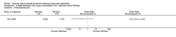
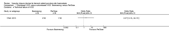
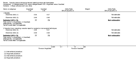
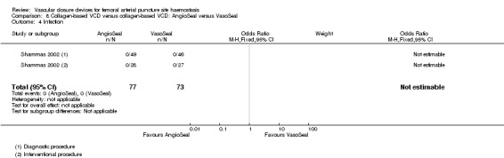
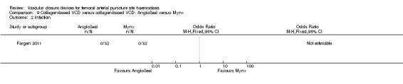
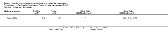
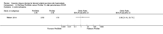
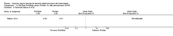
Main results: We included 52 studies (19,192 participants) in the review. We found studies comparing VCDs with extrinsic compression (sheath size ≤ 9 Fr), different VCDs with each other after endovascular (EVAR) and percutaneous EVAR procedures and VCDs with surgical closure after open exposure of the artery (sheath size ≥ 10 Fr). For primary outcomes, we assigned the quality of evidence according to GRADE (Grades of Recommendation, Assessment, Development and Evaluation) criteria as low because of serious imprecision and for secondary outcomes as moderate for precision, consistency and directness.For time to haemostasis, studies comparing collagen-based VCDs and extrinsic compression were too heterogenous to be combined. However, both metal clip-based (MD -14.81 minutes, 95% CI -16.98 to -12.63 minutes; five studies; 1665 participants) and suture-based VCDs (MD -14.58 minutes, 95% CI -16.85 to -12.32 minutes; seven studies; 1664 participants) were associated with reduced time to haemostasis when compared with extrinsic compression.For time to mobilisation, studies comparing collagen-, metal clip- and suture-based devices with extrinsic compression were too heterogeneous to be combined. No deaths were reported in the studies comparing collagen-based, metal clip-based or suture-based VCDs with extrinsic compression. For vascular injury requiring repair, meta-analyses demonstrated that neither collagen (OR 2.81, 95% CI 0.47 to 16.79; six studies; 5731 participants) nor metal clip-based VCDs (OR 0.49, 95% CI 0.03 to 7.95; three studies; 783 participants) were more effective than extrinsic compression. No cases of vascular injury required repair in the study testing suture-based VCD with extrinsic compression.Investigators reported no differences in the incidence of infection between collagen-based (OR 2.14, 95% CI 0.88 to 5.22; nine studies; 7616 participants) or suture-based VCDs (OR 1.66, 95% CI 0.22 to 12.71; three studies; 750 participants) and extrinsic compression. No cases of infection were observed in studies testing suture-based VCD versus extrinsic compression. The incidence of groin haematoma was lower with collagen-based VCDs than with extrinsic compression (OR 0.46, 95% CI 0.40 to 0.54; 25 studies; 10,247 participants), but no difference was evident when metal clip-based (OR 0.79, 95% CI 0.46 to 1.34; four studies; 1523 participants) or suture-based VCDs (OR 0.65, 95% CI 0.41 to 1.02; six studies; 1350 participants) were compared with extrinsic compression. The incidence of pseudoaneurysm was lower with collagen-based devices than with extrinsic compression (OR 0.74, 95% CI 0.55 to 0.99; 21 studies; 9342 participants), but no difference was noted when metal clip-based (OR 0.76, 95% CI 0.20 to 2.89; six studies; 1966 participants) or suture-based VCDs (OR 0.79, 95% CI 0.25 to 2.53; six studies; 1527 participants) were compared with extrinsic compression. For other adverse events, researchers reported no differences between collagen-based, clip-based or suture-based VCDs and extrinsic compression.Limited data were obtained when VCDs were compared with each other. Results of one study showed that metal clip-based VCDs were associated with shorter time to haemostasis (MD -2.24 minutes, 95% CI -2.54 to -1.94 minutes; 469 participants) and shorter time to mobilisation (MD -0.30 hours, 95% CI -0.59 to -0.01 hours; 469 participants) than suture-based devices. Few studies measured (major) adverse events, and those that did found no cases or no differences between VCDs.Percutaneous EVAR procedures revealed no differences in time to haemostasis (MD -3.20 minutes, 95% CI -10.23 to 3.83 minutes; one study; 101 participants), time to mobilisation (MD 1.00 hours, 95% CI -2.20 to 4.20 hours; one study; 101 participants) or major adverse events between PerClose and ProGlide. When compared with sutures after open exposure, VCD was associated with shorter time to haemostasis (MD -11.58 minutes, 95% CI -18.85 to -4.31 minutes; one study; 151 participants) but no difference in time to mobilisation (MD -2.50 hours, 95% CI -7.21 to 2.21 hours; one study; 151 participants) or incidence of major adverse events.
Authors' conclusions: For time to haemostasis, studies comparing collagen-based VCDs and extrinsic compression were too heterogeneous to be combined. However, both metal clip-based and suture-based VCDs were associated with reduced time to haemostasis when compared with extrinsic compression. For time to mobilisation, studies comparing VCDs with extrinsic compression were too heterogeneous to be combined. No difference was demonstrated in the incidence of vascular injury or mortality when VCDs were compared with extrinsic compression. No difference was demonstrated in the efficacy or safety of VCDs with different mechanisms of action. Further work is necessary to evaluate the efficacy of devices currently in use and to compare these with one other and extrinsic compression with respect to clearly defined outcome measures.
Conflict of interest statement
LR: none known. AA: none known. FC: travel/accommodation/meeting expenses: Gore Medical training (product training/aortic intervention, October 2014), Cook Medical training (Vista Education programme) (product training in embolisation, September 2014), Medtronic Aortic University (aortic stent grafting training, 2015), Vascutek (Terumo) (sponsorship for BSIR meeting, November 2014; sponsorship for LINC meeting, January 2015). RJ: none known.
Figures


Update of
References
References to studies included in this review
Amin 2000 {published data only}
-
- Amin FR, Yousufuddin M, Stbales R, Shamim Q, Al‐Nasser FM, Coats AJS, et al. Femoral haemostasis after transcatheter therapeutic intervention: a prospective randomised study of the AngioSeal device vs. the FemoStop device. International Journal of Cardiology 2000;76:235‐40. - PubMed
Amine 1999 {published data only}
-
- Amine el S, Teiger E, Aptecar E, Dubois‐Randé J‐L, Pernès J‐M, Dupouy P. Suture percutanée du point de ponction artériel fémoral après coronarographie diagnostique. Archives des Malaides du Coeur et des Vaisseaux 1999;92(11):1447‐53. - PubMed
Ansel 2006 {published data only}
-
- Ansel G, Yakubov S, Neilson C, Allie D, Stoler R, Hall P, et al. Safety and efficacy of staple‐mediated femoral arteriotomy closure: results from a randomised multicentre study. Catheterization and Cardiovascular Interventions 2006;67:546‐53. - PubMed
Behan 2007 {published data only}
-
- Behan MWH, Large JK, Patel NR, Lloyd GW, Sulke AN. A randomised controlled trial comparing the routine use of an AngioSeal STS device strategy with conventional femoral haemostasis methods in a district general hospital. International Journal of Clinical Practice 2007;61(3):367‐72. - PubMed
Brachmann 1998 {published data only}
-
- Brachmann J, Asnsah M, Kosinksi EJ, Schuler GC. Imprved clinical effectiveness with a collagen vascular hemostasis device for shortened immobilization time following diagnostic angiography and percutaneous transluminal coronary angioplasty. The American Journal of Cardiology 1998;81(12):1502‐5. - PubMed
Camenzind 1994 {published data only}
-
- Camenzind E, Grossholz M, Urban P, Dorsaz PA, Didier D, Meier B. Collagen application versus manual compression: a prospective randomized trial for arterial puncture site closure after coronary angioplasty. Journal of the American College of Cardiology 1994;24(3):655‐62. - PubMed
Carere 2000 {published data only}
-
- Carere RG, Webb JG, Buller CEH, Wilson M, Rahman T, Spinelli J, et al. Suture closure of femoral arterial puncture sites after coronary angioplasty followed by same‐day discharge. American Heart Journal 2000;139:52‐8. - PubMed
Castañeda 2003 {published data only}
-
- Castañeda F, Swischuk JL, Smouse B, Brady T. Gelatin sponge closure device versus manual compression after peripheral arterial catheterization procedures. Journal of Vascular and Interventional Radiology 2003;14(12):1517‐23. - PubMed
Chen 2013 {published data only}
Deuling 2008 {published data only}
-
- Deuling JHH, Vermeulen RP, Anthonio RA, Heuvel AFM, Jaarsma T, Jessurun G, et al. Closure of the femoral artery after cardiac catheterization: a comparison of AngioSeal, StarClose and manual compression. Catheterization and Cardiovascular Interventions 2008;71(4):518‐23. - PubMed
Diaz 2001 {published data only}
-
- Diaz De La Llera LS, Fournier Andray JA. Early deambulation following cardiac catheterization by the use of 6 Fr Angio‐Seal, a new hemostatic percutaneous puncture closure device. Revista Espanola de Cardiologia 2001;54(12):1406‐10. - PubMed
Doneaux 2001 {published data only}
-
- Doneaux P, Gach O, Martinez CH, Legrand V. Femoral puncture closure devices versus manual compression after percutaneous coronary interventions. European Heart Journal 2001;22(Suppl):216.
Fargen 2011 {published data only}
-
- Fargen KM, Hoh BL, Mocco J. A prospective randomized single‐blind trial of patient comfort following vessel closure: extravascular synthetic sealant closure provides less pain than a self‐tightening suture vascular compression device. Journal of Neurointerventional Surgery 2011;3:219‐33. - PubMed
-
- NCT00998023. Patient comfort with vascular closure. http://clinicaltrials gov/show/NCT00998023 (accessed 26 October 2013).
Gerckens 1998 {published data only}
-
- Gerckens U, Cattelaens N, Lampe EG, Grube E. Management of arterial puncture site after catheterization procedures: evaluating a suture‐mediated closure device. American Journal of Cardiology 1999;83(12):1658‐63. - PubMed
-
- Gerckens U, Cattelaens N, Müller R, Lampe E‐G, Grube E. Perkutaner nahtverschluß von femoralarteriezugängen nach diagnostischen herzkatheterisierungen oder koronarinterventionen. Sicherheit and effekitivtät einer neuen verschlußtechnik arterieller punktionsstellen. Herz 1998;23(1):27‐34. - PubMed
Gwechenberger 1997 {published data only}
-
- Gwechenberger M, Katzenschlager R, Heinz G, Gottsauner‐Wolf M, Probst P. Use of a collagen plug versus manual compression for sealing arterial puncture site after cardiac catheterization. Angiology 1997;48(2):121‐6. - PubMed
Hattab 2012 {published data only}
-
- Hattab M, Hokin M, Carreira VB, Elhadad S. A randomized trial comparing two vascular closure devices: PROGLIDE and the novel EXOSEAL after percutaneous femoral procedures. Journal of the American College of Cardiology 2012;60(17 Suppl B):B112.
Hermanides 2010 {published data only}
-
- Hermanides RS, Ottervanger JP, Dambrink JH, Boer MJ, Hoorntje JC, Gosselink AT, et al. Closure device or manual compression in patients undergoing percutaneous coronary intervention: a randomized comparison. Journal of Invasive Cardiology 2010;22(12):562‐6. - PubMed
Hermiller 2005 {published data only}
-
- Hermiller J, Simonton C, Hinohara T, Lee D, Cannon L, Mooney M, et al. Clinical experience with a circumferential clip‐based vascular closure device in diagnostic catheterization. Journal of Invasive Cardiology 2005;17(10):504‐10. - PubMed
Hermiller 2006 {published data only}
-
- DiDonato K, Jaff M, Hermiller J, Simonton C, Hinohara T, Mooney M. 6 month results for the CLIP trial. Transcatheter Cardiovascular Therapeutics. 2006; Vol. Abstract 479.
-
- Hermiller JB, Simonton C, Hinohara T, Cannon L, Mooney M, O'Shaughnessy C, et al. The StarClose® vascular closure system: interventional results from the CLIP study. Catheterization and Cardiovascular Interventions 2006;68(5):677‐83. - PubMed
-
- Jaff MR, Hadley G, Hermiller JB, Simonton C, Hinohara T, Cannon L, et al. The safety and efficacy of the StarClose Vascular CLosure system: the ultrasound substudy of the CLIP study. Catheterization and Cardiovascular Interventions 2006;68(5):684‐9. - PubMed
Holm 2014 {published data only}
-
- Holm NR, Sindberg B, Schou M, Maeng M, Kaltoft A, Bottcher M, et al. Randomised comparison of manual compression and FemoSeal vascular closure device for closure after femoral artery access coronary angiography: the CLOSure dEvices Used in everyday Practice (CLOSE‐UP) study. Eurointervention 2014;10(2):183‐90. - PubMed
-
- Sindberg B, Schou M, Hansen L, Christiansen KJ, Jørgensen KS, Søloft M, et al. Pain and discomfort in closure of femoral access coronary angiography. The CLOSuredEvices Used in everyday Practice (CLOSE‐UP) pain substudy. European Journal of Cardiovascular Nursing 2014;13(3):221‐6. - PubMed
Jensen 2008 {published data only}
-
- Jensen J, Saleh N, Jensen U, Svane B, Jönsson A, Tornvall P. The inflammatory response to femoral arterial closure devices: a randomized comparison among FemoStop, AngioSeal, and PerClose. Cardiovascular and Interventional Radiology 2008;31(4):751‐5. - PubMed
Juergens 2004 {published data only}
-
- Juergens CP, Leung DYC, Crozier JA, Wong AM, Robinson JTC, Lo S, et al. Patient tolerance and resource utilisation associated with an arterial closure versus an external compression device after percutaneous coronary intervention. Catheterization and Cardiovascular Interventions 2004;63:166‐70. - PubMed
Kalsch 2008 {published data only}
-
- Kalsch HIM, Eggebrecht H, Mayringer S, Konorza T, Sievers B, Sack S, et al. Randomized comparison of effects of suture‐based and collagen‐based vascular closure devices on post‐procedural leg perfusion. Clinical Research in Cardiology 2008;97(1):43‐8. - PubMed
Kussmaul 1995 {published data only}
-
- Kussmaul WG, Buchbinder M, Whitlow PL, Heuser RR, King SB, Kent KM, et al. Rapid arterial hemostasis and decreased access site complications after cardiac catheterization and angioplasty: results of a randomized trial of a novel hemostatic device. Journal of the American College of Cardiology 1995;25(7):1685‐92. - PubMed
Legrand 2005 {published data only}
-
- Legrand V, Doneux P, Martinez C, Gach O, Bellekens M. Femoral access management: comparison between two different vascular closure devices after percutaneous coronary intervention. Acta Cardiologica 2005;60(5):482‐8. - PubMed
Machnik 2012 {published data only}
-
- Machnik R, Pleniążek, Musiałek P, Przewłocki T, Tekieli Ł, Trystuła M, et al. Control of local haemostasis with the AngioSeal® vascular closure device in peripheral endovascular interventions via 6‐9 F femoral artery access. Postępy w Kardiologii Interwencyjnej 2012;8(1):1‐7.
Magosaki 1999 {published data only}
-
- Magosaki N, Hosoda S, Kawagishi N, Yamaguchi T, Sakatani H, Haze K, et al. Efficacy of hemostatic puncture closing device for hemostasis and early ambulation after coronary angiography and angioplasty: results of a multicentre trial. Journal of Cardiology 1999;34:131‐8. - PubMed
Martin 2008 {published data only}
-
- Martin JL, Pratsos A, Magargee E, Mayhew K, Pensyl C, Nunn M, et al. A randomized trial comparing compression, PerClose ProGlideTM and AngioSeal VIPTM for arterial closure following percutaneous coronary intervention: the CAP trial. Catheterization and Cardiovascular Interventions 2008;71(1):1‐5. - PubMed
Michalis 2002 {published data only}
-
- Michalis LK, Rees MR, Patsouras D, Katsouras CS, Goudevenos J, Pappas S, et al. A prospective randomized trial comparing the safety and efficacy of three commercially available closure devices (AngioSeal, VasoSeal and Duett). Cardiovascular and Interventional Radiology 2002;25:423‐9. - PubMed
Nelson 2014 {published data only}
-
- Nelson PR, Kracjer MSZ, Kansal N, Rao V. A multicenter, randomized, controlled trial of totally percutaneous access versus open femoral exposure for endovascular aortic aneurysm repair (the PEVAR trial). Journal of Vascular Surgery 2014;59:1181‐94. - PubMed
Noguchi 2000 {published data only}
-
- Noguchi T, Miyazaki S, Yasuda S, Baba T, Sumida H, Morii I, et al. A randomised controlled trial of ProStar‐Plus for haemostasis in patients after coronary angioplasty. European Journal of Vascular and Endovascular Surgery 2000;19(5):451‐5. - PubMed
Park 2005 {published data only}
Perlowski 2011 {published data only}
-
- Perlowski A, Jaff M, O'Shaughnessy D, Brueggeman C, Rosenfield K. StarClose device closure of femoral arteriotomy site versus manual compression in patients with peripheral arterial disease: the CLIPIT trial. Journal of the American College of Cardiology 2011;57(14(Suppl 1)):E1982.
Rastan 2008 {published data only}
-
- Rastan A, Sixt S, Schwarzwälder U, Schwarz T, Frank U, Bürgelin K, et al. VIPER‐2: a prospective, randomized single‐centre comparison of 2 different closure devices with a hemostatic wound dressing for closure of femoral artery access sites. Journal of Endovascular Therapy 2008;15:83‐90. - PubMed
Reddy 2004 {published data only}
-
- Reddy BK, Brewster PS, Walsh T, Burket MW, Thomas WJ, Cooper CJ. Randomized comparison of rapid ambulation using radial, 4 French femoral access, or femoral access with AngioSeal closure. Catheterization and Cardiovascular Interventions 2004;62:143‐9. - PubMed
Rickli 2002 {published data only}
-
- Rickli H, Unterweger M, Sütsch G, Brunner‐La Rocca HP, Sagmeister M, Ammann P, et al. Comparison of costs and safety of a suture‐mediated closure device with conventional manual compression after coronary artery interventions. Catheterization and Cardiovascular Interventions 2002;57(3):297‐302. - PubMed
Sanborn 1993 {published data only}
-
- Sanborn TA, Gibbs HH, Brinker JA, Knopf WD, Kosinski EK, Roubin GS. A multicentre randomised trial comparing a percutaneous collagen haemostasis device with conventional manual compression after diagnostic angiography and angioplasty. Journal of the American College of Cardiology 1993;22(5):1273‐9. - PubMed
Schräder 1992 {published data only}
-
- Schräder R, Steinbacher ST, Kaltenbach M. Randomisierter vergleich zwischen kollagenpplikation und druckverband zum verschluß der arteriellen punktionsstelle nach koronarangiographie und koronardilatation. Zeitschrift fűr Kardiologie 1992;81:507‐11. - PubMed
Schulz‐Schüpke 2014 {published data only}
-
- Schulz‐Schüpke S, Helde S, Gewalt S, Ibrahim T, LinhardtM, Haas K, et al. Comparison of vascular closure devices vs manual compression after femoral artery puncture. The ISAR‐CLOSURE randomized clinical trial. Journal of the American Medical Association 2014;312(19):1981‐7. - PubMed
SEAL Trial Study Team {published data only}
-
- The SEAL Trial Study Team. Assessment of the safety and efficacy of the DUETT vascular hemostasis device: final results of the Safe and Effective Vascular Hemostasis (SEAL) trial. American Heart Journal 2002;143(4):612‐9. - PubMed
-
- Zhang Z, Mahoney EM, Gershony G, Ellis S, Saucedo JF, Talley JD, et al. Impact of the Duett sealing device on quality of life and hospitalization costs for coronary diagnostic and interventional procedures: results from the Study of Economic and Quality of Life substudy of the SEAL trial. American Heart Journal 2001;142:982‐8. - PubMed
Seidelin 1997 {published data only}
-
- Seidelin PH, Adelman AG. Mobilization within thirty minutes of elective diagnostic coronary angiography: a feasibility study using a hemostatic femoral puncture closure device. Journal of Interventional Cardiology 1997;10:409‐15.
Shammas 2002 {published data only}
-
- Shammas NW, Rajendran VR, Alldredge SG, Witcik WJ, Robken JA, Lewis JR, et al. Randomized comparison of VasoSeal and AngioSeal closure devices in patients undergoing coronary angiography and angioplasty. Catheterization and Cardiovascular Interventions 2002;55:421‐5. - PubMed
Silber 1998 {published data only}
-
- Silber S, Björvik A, Mühling H, Rösch A. Usefulness of collagen plugging with VasoSeal® after PTCA as compared to manual compression with identical sheath dwell times. Catheterization and Cardiovascular Diagnosis 1998;43:421‐7. - PubMed
Sun 2009 {published data only}
-
- Sun JJ, Zhang HT, Huang CC, Luo HL, Wang JH, Tan WJ, et al. Three kinds of vascular closure devices StarClose, PerClose and Boomerang in the femoral artery hemostasis applications. Journal of Clinical Rehabilitative Tissue Engineering Research 2009;13(13):2485‐90.
Tron 2003 {published data only}
-
- Tron C, Koning R, Eltchaninoff H, Douillet R, Chassaing S, Sanchez‐Giron C, et al. A randomized comparison of a percutaneous suture device versus manual compression for femoral artery hemostasis after PTCA. Journal of Interventional Cardiology 2003;16(3):217‐21. - PubMed
Upponi 2007 {published data only}
-
- Upponi SS, Ganeshan AG, Warakaulle DR, Phillips‐Hughes J, Boardman P, Uberoi R. AngioSeal versus manual compression for haemostasis following peripheral vascular diagnostic and interventional procedures ‐ A randomized controlled trial. European Journal of Radiology 2007;61(2):332‐4. - PubMed
Veasey 2008 {published data only}
-
- Veasey RA, Large JK, Silberbauer J, Paul G, Taggu Wm Ellery S, Rathore VS, et al. A randomised controlled trial comparing StarClose and AngioSeal vascular closure devices in a district general hospital. International Journal of Clinical Practice 2008;62(6):912‐8. - PubMed
von Hoch 1995 {published data only}
-
- Hoch F, Neumann F‐J, Theiss W, Kastrati A, Schömig A. Efficacy and safety of collagen implants for haemostasis of the vascular access site after coronary balloon angioplasty and coronary stent implantation: a randomized study. European Heart Journal 1995;16(5):640‐6. - PubMed
Ward 1998 {published data only}
-
- Ward SR, Casale P, Raymond R, Kussmaul WG, Simpfendorfer C for the Angio‐Seal Investigators. Efficacy and safety of a hemostatic puncture closure device with early ambulation after coronary angiography. American Journal of Cardiology 1998;81:569‐72. - PubMed
Wetter 2000 {published data only}
-
- Wetter DR, Rickli H, Smekal A, Amann FW. Early sheath removal after coronary artery interventions with use of a suture‐mediated closure device: clinical outcome and results of Doppler US evaluation. Journal of Vascular and Interventional Radiology 2000;11:1033‐7. - PubMed
Wong 2009 {published data only}
-
- Wong SC, Bachinsky W, Cambier P, Stoler R, Aji J, Rogers JH, et al. A randomized comparison of a novel bioabsorbable vascular closure device versus manual compression in the achievement of hemostasis after percutaneous femoral procedures. Journal of the American College of Cardiology: Cardiovascular Interventions 2009;2(8):785‐93. - PubMed
Yadav 2003 {published data only}
-
- Yadav JS, Ziada KM, Almany S, Davis TP, Castaneda F. Comparisons of the QuickSeal femoral arterial closure system with manual compression following diagnostic and interventional catheterization procedures. American Journal of Cardiology 2003;91:1463‐6. - PubMed
References to studies excluded from this review
Baim 2000 {published data only}
-
- Baim DS, Knopf WD, Hinohara T, Schwarten DE, Schatz RA, Pinkerton CA, et al. Suture‐mediated closure of the femoral access site after cardiac catheterization: results of the Suture To Ambulate aNd Discharge (STAND I and STAND II) trials. American Journal of Cardiology 2000;85(7):864‐9. - PubMed
Beyer‐Enke 1996 {published data only}
-
- Beyer‐Enke SA, Soldner J, Zeitler E. Immediate sealing of arterial puncture site following femoropopliteal angioplasty: a prospective randomized trial. Cardiovascular and Interventional Radiology 1996;19:406‐10. - PubMed
Chalmers 2007 {published data only}
-
- Chalmers R, Clement Darling R, Wingard JT, Chetter I, Cutler B, Kern JA, FIBRIN SEALANT PV Investigator Group. A prospective, randomized controlled trial comparing the effects of a fibrin sealant (EVICEL) versus manual compression on hemostatic effectiveness during vascular surgical procedures utilizing polytetrafluorethylene graft material. Transfusion 2007;47(11(Suppl)):22A.
Chevalier 2000 {published data only}
-
- Chevalier B, Henry M, Pillière R, Koning R, Lancelin B, Puel J, et al. Duplex scan assessment of femoral access for coronary stenting: compression versus AngioSeal comparison in the HEMOSTASE trial. European Heart Journal 2000;21(Abstract Supplement 642):3522.
-
- Chevalier B, Lancelin B, Koning R, Henry M, Gommeaux A, Pilliere R, et al. Effect of a closure device on complication rates in high‐local‐risk patients: results of a randomized multicenter trial. Catheterization and Cardiovascular Interventions 2003;58(3):285‐91. - PubMed
Jean‐Baptiste 2008 {published data only}
-
- Jean‐Baptiste E, Hassen‐Khodja R, Haudebourg P, Bouillanne PJ, Declemy S, Batt M. Percutaneous closure devices for endovascular repair of infrarenal abdominal aortic aneurysms: a prospective, non‐randomized comparative study. European Journal of Vascular and Endovascular Surgery 2008;35(4):422‐8. - PubMed
Kurşaklioĝlu 2008 {published data only}
-
- Kurşaklioĝlu H, Iyisoy A, Barҫin C, Celik T, Nitzan R, Köse S, et al. The experience with the Epiclose‐T vascular access closure device: a human study. Anadolu Kardiyoloju Dergisi 2008;8(1):38‐42. - PubMed
Larzon 2015 {published data only}
-
- Larzon T, Roos H, Gruber G, Henrikson O, Magnuson A, Falkenberg M. A randomised controlled trial of the fascia suture technique compared with a suture‐mediated closure device for femoral arterial closure after endovascular aortic repair. European Jouranl of Vascular and Endovascular Surgery 2015;49(2):166‐73. - PubMed
Leinbudgut 2013 {published data only}
-
- Leibundgut G, Pache J, Schulz S, Berger PB, Ferenc M, Gick M, et al. Collagen plug vascular closure devices and reduced risk of bleeding with bivalirudin versus heparin plus abciximab in patients undergoing percutaneous coronary intervention for non ST‐segment elevation myocardial infarction. Journal of Interventional Cardiology 2013;26(6):623‐9. - PubMed
Lupi 2012 {published data only}
-
- Lupi A, Rognoni A, Secco CG, Lazzero M, Plebani L, Cossa G. Different spectrum of vascular complications after AngioSeal deployment of manual compression. Journal of Invasive Cardiology 2012;24(3):90‐6. - PubMed
Neudecker 2003 {published data only}
-
- Neudecker A, Manke C, Lenhart M, Zorger N, Paetzel C, Feurbach S, et al. Evaluation of a haemostatic device with percutaneous collagen application (VasoSeal) compared to a mechanical compression system (Compressar) after transfemoral catheterization of patients suffering from arterial occlusive disease [Evaluation eines Verschlusssystems mit perkutaner Kollageneinbringung (VasoSeal) im Vergleich zu einem mechanischen Kompressionssystem (Compressar) nach Femoralispunktion bei Patienten mit AVK]. Rofo: Fortschritte auf dem Gebiete der Rontgenstrahlen und der Nuklearmedizin 2003;175(5):676‐81. - PubMed
Ratnam 2007 {published data only}
-
- Ratnam LA, Raja J, Munneke GJ, Morgan RA, Belli AM. Prospective non‐randomized trial of manual compression and AngioSeal and StarClose arterial closure devices in common femoral punctures. Cardiovascular and Interventional Radiology 2007;30(2):182‐8. - PubMed
Slaughter 1995 {published data only}
-
- Slaughter PM, Chetty R, Flintoft VF, Lewis S, Sykora K, Beattie DM, et al. A single center randomized trial assessing use of a vascular hemostasis device vs. conventional manual compression following PTCA: what are the potential resource savings?. Catheterization and Cardiovascular Diagnosis 1995;34(3):210‐4. - PubMed
Smilowitz 2012 {published data only}
-
- Smilowitz NR, Kirtane AJ, Guiry M, Gray WA, Dolcimascolo P, Querijero M, et al. Practices and complications of vascular closure devices and manual compression in patients undergoing elective transfemoral coronary procedures. American Journal of Cardiology 2012;110(2):177‐82. - PubMed
Starnes 2003 {published data only}
-
- Starnes BW, O'Donnell SD, Gillespie DL, Goff JM, Rosa P, Parker MV, et al. Percutaneous arterial closure in peripheral vascular disease: a prospective randomized evaluation of the PerClose device. Journal of Vascular Surgery 2003;38(2):263‐71. - PubMed
References to studies awaiting assessment
NCT01297322 {published data only}
-
- NCT01297322. RESPECT Trial ‐ (Rapid Extravascular Sealing Via PercutanEous Collagen ImplanT). http://clinicaltrials.gov/showNCT01297322 (accessed 26 October 2013).
References to ongoing studies
ACTRN12611001248954 {published data only}
-
- ACTRN12611001248954. Femoral artery closure using the Cardiva VASCADE Vascular Closure System (VCS) to reduce time to hemostasis and reduce time to ambulation and hospital discharge versus manual compression in patients who have undergone diagnostic or interventional endovascular catheterization procedures utilizing 6 Fr or 7 Fr procedural sheaths. http://www anzctr org au/ACTRN12611001248954 (accessed 26 October 2013).
CTRI/2014/09/004946 {published data only}
-
- CTRI/2014/09/004946. Impact of femoral access site closure after percutaneous coronary interventions. A comparison between manual compression and the use of vascular closure device (PerClose ProGlide). http://apps.who.int/trialsearch/Trial2.aspx?TrialID=CTRI/2014/09/004946 (accessed 27 May 2015).
DRKS00000802 {published data only}
-
- DRKS00000802. A prospective randomized trial to evaluate the complication rate and patient comfort using three different vascular closure strategies following coronary angiography. http://drks‐neu.uniklinik‐freiburg.de/drks_web/navigate.do?navigationId=trial.HTML&TRIAL_ID=DR... (accessed 26 October 2013).
NCT00264264 {published data only}
-
- NCT00264264. Arterial closure vs direct compression for hemostasis after PCI ‐ ACDC Trial. http://clinicaltrials.gov/show/NCT00264264 (accessed 25 October 2013).
NCT00428155 {published data only}
-
- NCT00428155. Arterial Closure Device Comparison Trial II ‐ ACDC Trial II. http://clinicaltrials.gov/show/NCT00428155 (accessed 25 October 2103).
NCT01389375 {published data only}
-
- NCT01389375. Instrumental sealing of arterial puncture site closure device versus manual compression trial (ISAR‐CLOSURE). http://clinicaltrials.gov/show/NCT01389375 (accessed 26 October 2013).
NCT01600482 {published data only}
-
- NCT01600482. Clinical investigation for safety and efficacy study of CELT ACD arterial closure device. http://clinicaltrials.gov/show/NCT01600482 (accessed 26 October 2013).
NCT01669382 {published data only}
-
- NCT01669382. AngioSeal® vs. ExoSeal® for closure of arterial puncture sites. http://clinicaltrials.gov/show/NCT01669382 (accessed 26 October 2013).
NCT02061696 {published data only}
-
- NCT02061696. A randomized controlled trial to assess safety and efficacy of AXERA (device name) 2 Access System compared to manual compression. https://clinicaltrials.gov/ct2/show/NCT02061696 (accessed 27 May 2015).
NCT02234830 {published data only}
-
- NCT02234830. Randomized comparison of ExoSeal® and AngioSeal vascular closure devices: the CLOSE‐UP II trial. https://clinicaltrials.gov/ct2/show/NCT02234830 (accessed 27 May 2015).
NCT02237430 {published data only}
-
- NCT02237430. Randomized comparison of MynxGrip vascular closure device and manual compression for closure after femoral access angiography. The Closure Devices Used in Every Day Practice Study, CLOSE‐UP III Trial. https://clinicaltrials.gov/ct2/show/NCT02237430 (accessed 27 May 2015). - PMC - PubMed
Additional references
Bechara 2010
-
- Bechara CF, Annambhotla S, Lin PH. Access site management with vascular closure devices for percutaneous transarterial procedures. Journal of Vascular Surgery 2010;52(6):1682‐96. - PubMed
Biancari 2010
-
- Biancari F, D'Andrea V, Marco C, Savino G, Tiozzo V, Catania A. Meta‐analysis of randomized trials on the efficacy of vascular closure devices after diagnostic angiography and angioplasty. American Heart Journal 2010;159:518‐31. - PubMed
GRADE 2004
Higgins 2011
-
- Higgins JPT, Green S (editors). Cochrane Handbook for Systematic Reviews of Interventions Version 5.1.0 [updated March 2011]. The Cochrane Collaboration, 2011. www.cochrane‐handbook.org.
Jiang 2015
Koreny 2004
-
- Koreny M, Riedmüller E, Nikfardjam M, Siostrzonek P, Müllener M. Arterial puncture closing devices compared with standard manual compression after cardiac catheterization. Systematic review and meta‐analysis. Journal of American Medicine Association 2004;291(3):350‐7. - PubMed
Kurşaklioğlu 2008
-
- Kurşaklioğlu H, Iyisoy A, Barçin C, Celik T, Nitzan R, Köse S, et al. The experience with the Epiclose‐T vascular access closure device: a human study. Anadolu Kardiyoloji Dergisi ‐ The Anatolian Journal of Cardiology 2008;8(1):38‐42. - PubMed
Merriweather 2012
-
- Merriweather N, Sulzbach‐Hoke LM. Managing risk of complications at femoral vascular access sites in percutaneous coronary intervention. Critical Care Nurse 2012;32(5):16‐30. - PubMed
Nikolsky 2004
-
- Nikolsky E, Mehran R, Halkin A, Aymong ED, Mintz GS, Lasic Z, et al. Vascular complications associated with arteriotomy closure devices in patients undergoing percutaneous coronary procedures: a meta‐analysis. Journal of the American College of Cardiology 2004;44(6):1200‐9. - PubMed
Scheinert 2007
-
- Scheinert D, Sievert H, Turco MA, Schmidt A, Hauptmann KE, Mueller R, et al. The safety and efficacy of an extravascular, water‐soluble sealant for vascular closure: initial clinical results for Mynx. Catheterization and Cardiovascular Interventions 2007;70(5):627‐33. - PubMed
Schwartz 2010
-
- Schwartz BG, Burstein S, Economides C, Kloner RA, Shavelle DM, Mayeda GS. Review of vascular closure devices. Journal of Invasive Cardiology 2010;22(12):599‐607. - PubMed
Sterne 2001
-
- Sterne JA, Egger M. Funnel plots for detecting bias in meta‐analysis: guidelines on choice of axis. Journal of Clinical Epidemiology 2001;54(10):1046‐55. - PubMed
Zahn 1997
-
- Zahn R, Fromm E, Thoma S, Lotter R, Zander M, Wagner S. Local venous thrombosis after cardiac catheterization. Angiology 1997;48:1‐7. - PubMed
Publication types
MeSH terms
Substances
Grants and funding
LinkOut - more resources
Full Text Sources
Other Literature Sources
Miscellaneous