. 2016 Mar 7:7:10918.
doi: 10.1038/ncomms10918.

Increased amyloidogenic APP processing in APOE ɛ4-negative individuals with cerebral β-amyloidosis

Affiliations

Increased amyloidogenic APP processing in APOE ɛ4-negative individuals with cerebral β-amyloidosis

Niklas Mattsson et al. Nat Commun. .

Abstract

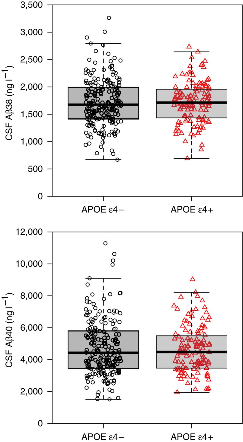
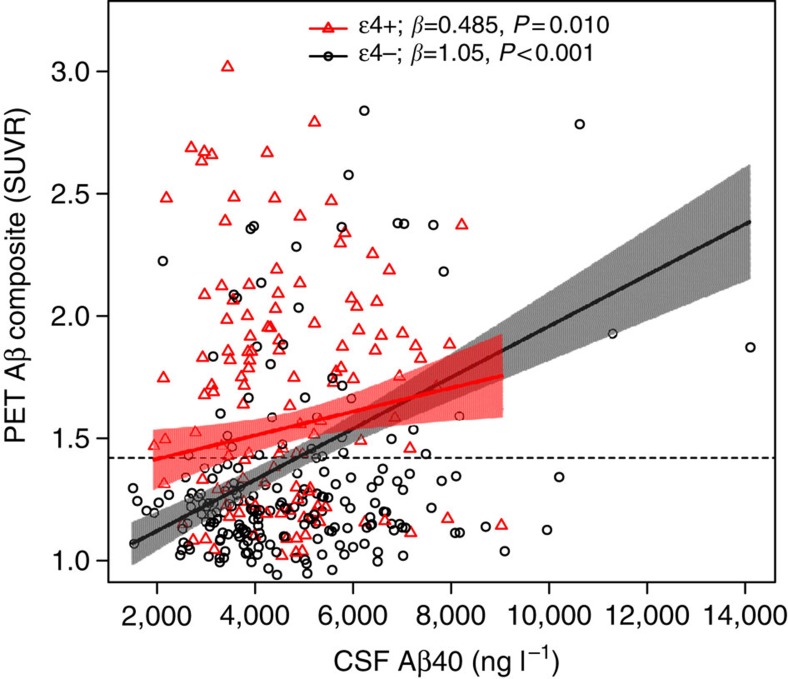
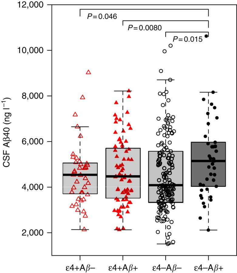
Increased APP (amyloid precursor protein) processing causes β-amyloid (Aβ) accumulation in autosomal dominant Alzheimer's disease (AD), but it is unclear if it also affects sporadic Aβ accumulation. We tested healthy controls and patients with mild cognitive symptoms (N=331) in the BioFINDER study, using cerebrospinal fluid (CSF) Aβ40 as a surrogate for amyloidogenic APP processing. We find that levels of brain Aβ fibrils (measured by 18F-flutemetamol PET) are independently associated with high CSF Aβ40 (P<0.001) and APOE ɛ4 (P<0.001). The association between CSF Aβ40 and brain Aβ is stronger in APOE ɛ4-negative than in positive people (P=0.0080). The results are similar for CSF Aβ38 and for a combination of CSF Aβ38 and CSF Aβ40. In conclusion, sporadic Aβ accumulation may be partly associated with increased amyloidogenic APP production, especially in APOE ɛ4-negative subjects. The risk for sporadic AD may consequently depend on increased Aβ production, in addition to decreased Aβ clearance.


Conflict of interest statement

N.M., P.S.I., S.P., E.S., L.H. and O.H. report no conflicts of interest. K.B. has served as a consultant for Eli Lilly, Novartis, Roche Diagnostics and Sanofi-Aventis, and at Advisory Boards for IBL International and lecturing for Fujirebio Europe and Lundbeck. K.B. and H.Z. are co-founders of Brain Biomarker Solutions in Gothenburg AB, a GU Holding-based platform company at the University of Gothenburg.

Figures

Figure 1
Figure 1. CSF Aβ38 and Aβ40 in APOE ɛ4- and APOE ɛ4+ subjects.
Observed data for CSF Aβ38 and CSF Aβ40 by APOE ɛ4 status. The individual observations are overlaid on boxplots (thick lines are medians, box limits are 25th and 75th percentiles). APOE ɛ4 did not affect levels of CSF Aβ38 (Mann–Whitney U-test, P=0.75; t-test, P=0.95; linear regression adjusted for age, sex and diagnosis, P=0.99) or CSF Aβ40 (Mann–Whitney U-test, P=0.84; t-test, P=0.43; linear regression adjusted for age, sex and diagnosis, P=0.26). This supports the notion that CSF Aβ38 and Aβ40 are unaffected by APOE ɛ4-mediated changes in Aβ clearance.
Figure 2
Figure 2. PET Aβ as a function of CSF Aβ40 and APOE ɛ4.
Observed PET and CSF Aβ40 data. Slopes are modelled from a linear regression adjusted for CSF Aβ42, sex, age and diagnostic group. The shaded areas indicate 95% confidence intervals for the slopes. The dotted line indicates a cutoff for clinically significant PET Aβ load (1.42 SUVR). β-coefficients (divided by 10−4) and P values for the slopes within APOE ɛ4-positive and separately for APOE ɛ4-negative subjects are shown in the legend. The interaction between CSF Aβ40 and APOE ɛ4 is significant, indicating that the correlation between CSF Aβ40 and PET Aβ differs by APOE ɛ4 status (P=0.0080). The results did not change significantly when removing outliers (CSF Aβ40>10,000 ng l−1).
Figure 3
Figure 3. CSF Aβ40 in different combinations of PET Aβ and APOE ɛ4.
Observed CSF Aβ40 for different combinations of APOE ɛ4 and PET Aβ positivity and negativity. The individual observations are overlaid on boxplots (thick lines are medians, box limits are 25th and 75th percentiles). CSF Aβ40 was significantly increased in the PET Aβ+ & APOE ɛ4− group compared with PET Aβ- & APOE ɛ4−, which was the reference category (P=0.015, using linear regression adjusted for age, sex, diagnosis and WML). No other group had significant different CSF Aβ40 compared with the PET Aβ- & APOE ɛ4− group (P=0.62–0.90). The groups were PET Aβ- & APOE ɛ4−, N=158; PET Aβ- & APOE ɛ4+, N=39; PET Aβ+ & APOE ɛ4−, N=41; and PET Aβ+ & APOE ɛ4+, N=75. The total N=313 for this analysis was smaller than the total study population (N=331) due to missing data for WML in 18 subjects (but the main results did not differ when WML was not included and the analysis was done on the whole study population). One data point is excluded from the graph for visual clarity (CSF Aβ40 14110, ng l−1, PET Aβ+ & APOE ɛ4−).

References

    1. Bateman R. J. et al. Clinical and biomarker changes in dominantly inherited Alzheimer's disease. N. Engl. J. Med. 367, 795–804 (2012). - PMC - PubMed
    1. Villemagne V. L. et al. Amyloid β deposition, neurodegeneration, and cognitive decline in sporadic Alzheimer's disease: a prospective cohort study. Lancet Neurol. 12, 357–367 (2013). - PubMed
    1. Jansen W. J. et al. Prevalence of cerebral amyloid pathology in persons without dementia: a meta-analysis. JAMA. 313, 1924–1938 (2015). - PMC - PubMed
    1. Klunk W. E. et al. Imaging brain amyloid in Alzheimer's disease with Pittsburgh Compound-B. Ann. Neurol. 55, 306–319 (2004). - PubMed
    1. Blennow K., Mattsson N., Schöll M., Hansson O. & Zetterberg H. Amyloid biomarkers in Alzheimer's disease. Trends Pharmacol. Sci. 36, 297–309 (2015). - PubMed

Publication types

Substances