. 2016 Mar 7:7:10738.
doi: 10.1038/ncomms10738.

Catch-bond mechanism of the bacterial adhesin FimH

Affiliations

Catch-bond mechanism of the bacterial adhesin FimH

Maximilian M Sauer et al. Nat Commun. .

Abstract

Ligand-receptor interactions that are reinforced by mechanical stress, so-called catch-bonds, play a major role in cell-cell adhesion. They critically contribute to widespread urinary tract infections by pathogenic Escherichia coli strains. These pathogens attach to host epithelia via the adhesin FimH, a two-domain protein at the tip of type I pili recognizing terminal mannoses on epithelial glycoproteins. Here we establish peptide-complemented FimH as a model system for fimbrial FimH function. We reveal a three-state mechanism of FimH catch-bond formation based on crystal structures of all states, kinetic analysis of ligand interaction and molecular dynamics simulations. In the absence of tensile force, the FimH pilin domain allosterically accelerates spontaneous ligand dissociation from the FimH lectin domain by 100,000-fold, resulting in weak affinity. Separation of the FimH domains under stress abolishes allosteric interplay and increases the affinity of the lectin domain. Cell tracking demonstrates that rapid ligand dissociation from FimH supports motility of piliated E. coli on mannosylated surfaces in the absence of shear force.


Figures

Figure 1
Figure 1. FimH·DsG resembles fimbrial tip FimH.
(a) Preparation of the FimH·DsG complex by DSE. Left: reaction scheme of the DSE reaction, in which DsG displaces the FimC chaperone from the FimH pilin domain. Right: kinetics of the FimH·DsG complex formation at 37 °C, monitored by analytical gel filtration. DSE was initiated by mixing the FimC·FimH complex (15 μM) with excess DsG peptide (50 μM). Samples were removed after different incubation times, rapidly cooled on ice and immediately subjected to gel filtration. The reaction can be followed by the decrease in the FimC·FimH complex concentration and the simultaneous increase in the concentrations of FimH·DsG and free FimC (FimC and FimH·DsG coelute as a single peak at ∼12 ml). The chromatogram at the bottom shows that the FimC·FimH complex is stable against dissociation/aggregation under the chosen conditions. The rate constant of DSE estimated from these data is ∼0.5 M−1 s−1. (b) Structure of FimHF18·DsG (lectin domain FimHL, red; pilin domain FimHP, yellow; DsG, blue; circle and square indicate N- and C termini, respectively). (c) FimH from the fimbrial tip structure (left, PDB ID: 3JWN (ref. 17); FimG, blue; FimF, green) is superposed onto FimHF18·DsG based on their pilin domains (aa. 160–279), in the superposition (right) fimbrial FimH is shown in grey. (d) Close-up on the DsG peptide (stick representation) bound to FimHF18·DsG with 2FoFc electron density map. Backbone hydrogen bonds of the DsG peptide and β-strands 2 (β2) and 9 (β9) of FimHP are indicated.
Figure 2
Figure 2. Crystallographic analysis of FimH conformational states.
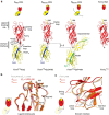
(a) FimHF18·DsG in the Afree (left) and in the Abound states (FimHF18·DsG·HM) in comparison with the Sbound state of FimHK12·DsF·HM and the isolated FimHLF18·HM (right). The FimHL, FimHP, DsF and DsG are coloured in red, yellow, green and blue. The experimentally in crystallo trapped orientation of FimHP in FimHK12·DsF·HM and a modelled position based on a hinge motion stretching around Gly157 is indicated. A schematic representation for each crystal structure, similar to Fig. 1a, is given. The tip of the clamp loop and the C terminus of FimHL are indicated as a circle and diamond, respectively. (b) Comparison of the conformation of the ligand-binding site in the Abound (red) and Sbound (orange) states with the isolated lectin domain FimHL (grey) and (c) comparison of the interdomain interface of the lectin domain.
Figure 3
Figure 3. The interdomain region in the Sbound state.
Close-up of the interdomain region of FimHK12·DsF·HM in the Abound form (left) and FimHK12·DsF·HM in the Sbound state (right). A cartoon representation for each crystal structure, similar to Fig. 1a, is given. Key residues in the interface are shown as sticks. FimHP, FimHL and DsF are coloured in yellow, red and green, respectively.
Figure 4
Figure 4. Kinetics of HM binding and release by full-length FimH.
(a) Fluorescence spectra (excitation at 280 nm) of FimHLF18 (2 μM; red lines) and FimHF18·DsG (2 μM; black lines) in the absence (solid lines) or presence of 200 μM HM (dotted lines). (b) Equilibrium titration of FimHF18·DsG (2 μM) with HM, recorded via the fluorescence increase at 320 nm. The total concentration of HM is plotted against the recorded fluorescence signal. Data were fitted (solid line) according to equation (2) (cf. experimental section) and yielded a Kd value of 9.9±1.5 μM. (c) Stopped-flow fluorescence kinetics of HM binding to FimHF18·DsG (1.0 μM), recorded via the fluorescence change above 320 nm. The HM concentration was varied between 0 and 50 μM. Five representative traces are shown (HM concentrations are given in μM). The fluorescence traces were globally fitted according to a second-order binding and first-order dissociation reaction (solid lines; Table 2). (d) Amplitudes of the reactions monitored in c, plotted against the total HM concentration. Data were fitted (solid line) according to equation (2), yielding a Kd value of 12±1 μM.
Figure 5
Figure 5. HM binding and release by the isolated FimH lectin domain FimHL.
Analysis of FimHL·HM interactions based on competition between HM and the synthetic fluorescent GN-FP-4 ligand. (a) HM binding to FimHL analysed by displacement of GN-FP-4 from FimHL variants as indicated. An equimolar mixture of FimHL and GN-FP-4 (1 μM each) was incubated with different HM concentrations (10 nM–3.2 mM) for >18 h. GN-FP-4 displacement is monitored by a decrease in fluorescence polarization at 528±20 nm (excitation at 485 nm). Data were fitted (solid lines) according to a mechanism in which two ligands compete for the same binding site, with fixed Kd values for GN-FP-4 binding (cf. Table 2). (b) Kinetics of HM dissociation from FimHL. A solution with equimolar concentrations of FimHL and HM (3 μM each, guaranteeing >95% occupancy with HM) was mixed with excess GN-FP-4 (10 μM), and the decrease in GN-FP-4 fluorescence at 520 nm as a consequence of HM dissociation and GN-FP-4 binding was recorded (Supplementary Fig. 5f–j). The obtained first-order kinetics are independent of the GN-FP-4 concentration and thus directly monitor HM dissociation.
Figure 6
Figure 6. Cell-tracking analysis of bacterial motility on mannosylated surfaces.
E. coli cells piliated with FimHF18 or the FimHF18-Ala188Asp variants were tracked under static conditions in the absence of shear force. (a) The fraction of bacteria attached to mannose-coated (1M-BSA) or BSA-coated surface (negative control) at the beginning of the time-lapse movies (white bars) and after 5 min (black bars) are given. Bacterial motility on 1M-BSA was analysed in the absence and presence of HM. The delay between application of bacteria and movie recording was ∼1 min. (b) Fraction of tracked cells that were pre-attached (yellow; speed <0.5 μm s−1), permanently attach (red), were mobile (white), transiently attach (green) or permanently detach (blue) during the entire observation time (5 min). Right: schematic depiction of the observed cell behaviour. FimHF18-piliated E. coli show almost exclusively transient attachment events on 1M-BSA. FimHF18-Ala188Asp-piliated E. coli show less transient attachment but enhanced permanent attachment to 1M-BSA. Transient and permanent attachment to 1M-BSA is significantly reduced in the presence of HM. For each experiment five to seven independent replicates were analysed.
Figure 7
Figure 7. Catch-bond mechanism of FimH-mediated cell adhesion.
(a) In the absence of tensile mechanical force, formation of the FimH-Uroplakin 1a (UPIa) complex comprises the highly dynamic transition of the Afree to the Abound state. The reaction likely proceeds via a transient encounter complex (indicated in square brackets). The reaction of the encounter complex to Abound is not rate-limiting and must have a half-life of less than 1 ms. Dissociation of the receptor from the FimH lectin domain in the Abound state is promoted via dynamic allostery by the pilin domain that acts as a negative allosteric regulator. The reaction from Abound to the encounter complex corresponds to koff. Fast binding and release of UPIa by FimH enables bacterial motility on the bladder epithelium. (b) Shear force increases the population of the Sbound state of FimH, in which the pilin and lectin domains are separated. The dissociation of Sbound under shear force is slowed down up to 100,000-fold compared with Abound. The indicated rate constants and half-lives correspond to the interaction between FimHF18 and the model ligand HM. Rate limiting reactions are indicated by solid arrows, and fast, non-limiting reactions by dashed arrows.

References

    1. Marshall B. T. et al. Direct observation of catch bonds involving cell-adhesion molecules. Nature 423, 190–193 (2003). - PubMed
    1. Evans E., Leung A., Heinrich V. & Zhu C. Mechanical switching and coupling between two dissociation pathways in a P-selectin adhesion bond. Proc. Natl Acad. Sci. USA 101, 11281–11286 (2004). - PMC - PubMed
    1. Buckley C. D. et al. Cell adhesion. The minimal cadherin-catenin complex binds to actin filaments under force. Science 346, 1254211 (2014). - PMC - PubMed
    1. Akiyoshi B. et al. Tension directly stabilizes reconstituted kinetochore-microtubule attachments. Nature 468, 576–579 (2010). - PMC - PubMed
    1. Fiore V. F., Ju L., Chen Y., Zhu C. & Barker T. H. Dynamic catch of a Thy-1-alpha5beta1+syndecan-4 trimolecular complex. Nat. Commun. 5, 4886 (2014). - PubMed

Publication types

MeSH terms