. 2016 Mar 16;89(6):1173-1179.
doi: 10.1016/j.neuron.2016.02.001. Epub 2016 Mar 3.

Thalamocortical Projections onto Behaviorally Relevant Neurons Exhibit Plasticity during Adult Motor Learning

Affiliations

Thalamocortical Projections onto Behaviorally Relevant Neurons Exhibit Plasticity during Adult Motor Learning

Jeremy S Biane et al. Neuron. .

Abstract

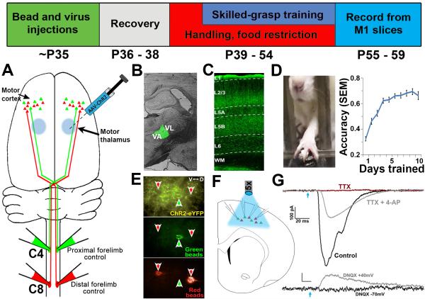
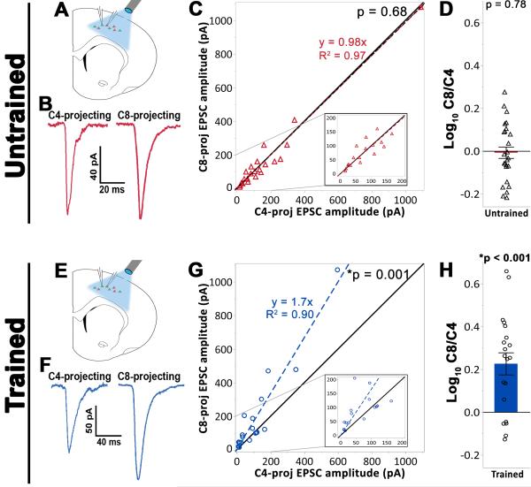
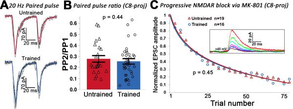
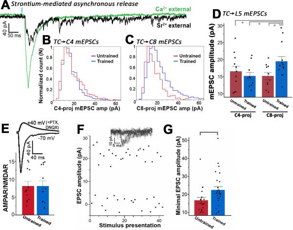
Layer 5 neurons of the neocortex receive direct and relatively strong input from the thalamus. However, the intralaminar distribution of these inputs and their capacity for plasticity in adult animals are largely unknown. In slices of the primary motor cortex (M1), we simultaneously recorded from pairs of corticospinal neurons associated with control of distinct motor outputs: distal forelimb versus proximal forelimb. Activation of ChR2-expressing thalamocortical afferents in M1 before motor learning produced equivalent responses in monosynaptic excitation of neurons controlling the distal and proximal forelimb, suggesting balanced thalamic input at baseline. Following skilled grasp training, however, thalamocortical input shifted to bias activation of corticospinal neurons associated with control of the distal forelimb. This increase was associated with a cell-specific increase in mEPSC amplitude but not presynaptic release probability. These findings demonstrate distinct and highly segregated plasticity of thalamocortical projections during adult learning.


Figures

Figure 1
Figure 1. Experimental overview
Top: Timeline of experiments. (A) Retrograde tracer injections at levels C4 and C8 of the spinal cord label distinct corticospinal projection populations originating in layer 5 of M1. (B) To selectively express ChR2 in motor thalamic nuclei, AAV-ChR2 was targeted to the VA/VL complex of the thalamus. (C) TC axonal expression of ChR2-EYFP was robust in layer 5 (the location of corticospinal cell bodies) of the caudal forelimb region of M1. (D) Skilled grasp training was conducted over 10 days, leading to a significant increase in pellet retrieval accuracy (repeated-measures ANOVA, p < 0.001). (E) Following the completion of training, slices of M1 were prepared and C4- and C8-projecting corticospinal neurons were targeted for simultaneous whole-cell recording. Disambiguation of eYFP signal from green bead fluorescence was achieved via a narrow band GFP filter. (F) Photostimulation at ~470nm was applied across the cortical slice via 5x objective to selectively stimulate thalamocortical axons. (G) Corticospinal neurons receive direct monosynaptic input from TC axons; upper traces: photo-induced EPSCs were abolished in the presence of 1μM TTX (red trace), but rescued following addition of 100 μM 4-AP (grey trace); lower traces: the presence of the AMPAR antagonist DNQX abolished EPSCs when neurons were held at −70mV (black trace). Releasing the Mg2+ block from NMDA channels via depolarization to +40mV unmasked monosynaptic NMDA-mediated currents (grey trace). Blue arrow = light onset.
Figure 2
Figure 2. Plasticity of thalamocortical projections onto trained, grasp-related C8-projecting layer 5 corticospinal neuronal subpopulations
(A) – (D) Experimental results from baseline (untrained) animals. (A) Neighboring C4- and C8-projecting cell pairs were targeted for whole-cell patch clamp in slices containing M1. (B) Sample bulk-stimulation EPSCs from a simultaneously patched C4- and C8-projecting cell pair demonstrating comparable evoked response amplitude in both neuronal subtypes. (C) Analysis across all cell pairs indicated that thalamocortical input was balanced across corticospinal subpopulations under baseline conditions (Wilcoxon signed-rank test against unity, p = 0.95). Black line = unity. Colored dashed line = linear fit of data. Inset: magnified view for smaller amplitude responses. (D) The log10 of the ratio of C8-projecting to C4-projecting EPSC peak amplitudes. Individual data points represent the average of all cell pairs recorded in a single slice (see methods). Under baseline conditions, EPSC amplitude did not differ across corticospinal subpopulations (one-sample t-test against 0, p = 0.78). (E) – (H) Experimental results from trained animals. (E) The recording setup was the same as for untrained animals. (F) Sample bulk stimulation EPSCs from a simultaneously patched cell pair indicating a larger evoked response in the C8-projecting cell relative to the C4-proceting cell. (G) Analysis across all cell pairs indicated that skilled motor learning is associated with a greater thalamocortical drive onto the C8-vs C4-projecting cell population (Wilcoxon signed-rank test against unity, p < 0.001). (H) The ratio of C8-projecting:C4-projecting EPSC peak amplitudes (C8/C4) following training deviated significantly from unity (one sample t-test against 0, p < 0.001), and was also significantly greater than that observed in untrained animals ((D) vs (H); (Welch’s unequal variances t-test, p < 0.001).
Figure 3
Figure 3. Presynaptic release probability is unaffected by skilled motor training
(A) Postsynaptic responses to a paired light pulse in C8-projecting neurons from an untrained (upper) or trained (lower) animal. Black traces = individual responses. Colored traces = averaged response. (B) The average paired-pulse ratio did not differ across training conditions for either C8-projecting or C4-projecting cells (C8: t-test on log transformed data (see methods), p = 0.44; C4: t-test on log transformed data, p = 0.36; C4-projecting data not shown). (C) The NMDAR-mediated EPSC declined at a similar rate in the C8-projecting subpopulation in trained and untrained animals (t-test, p = 0.45). Inset: progressive decline of the NMDAR-mediated response from a sample neuron. All cells were held at +40mV in the presence of 20 μM MK-801, 20 μM DNQX, and 20 μM picrotoxin.
Figure 4
Figure 4. Postsynaptic quantal amplitude increases selectively onto the C8-projecting subpopulation following skilled motor training
(A) Sample postsynaptic responses to photostimulation (blue bar) of TC axons. Green trace = external solution containing 2.5 mM calcium. Black trace = same cell after calcium was replaced with 3 mM strontium. Note the diminished synchronous release in the presence of strontium, and the presence of individual release events that persist for 500+ ms following photostimulation. (B) The mEPSC amplitude distribution did not differ across training conditions in the C4-projecting population (K-S test, p = 0.6), (C) but the frequency of larger responses increased significantly with training in the C8-projecting population (K-S test, p < 0.01). (D) The mean mEPSC amplitude for trained C8-projecting cells was also significantly greater compared to all other conditions (Wilcoxon test, p< 0.05). (E) Upper trace: NMDAR-mediated EPSC with cell held at +40mV in the presence of DNQX and PTX. Lower trace: AMPAR-mediated response with cell held at −70mV. The AMPAR/NMDAR ratio for the TC✧C8 pathway was unaffected by training (t-test, p = 0.99). (F) Top traces: superimposed postsynaptic responses to light presentation at the minimal stimulation amplitude. Main: minimal optical stimulation parameters resulted in a ~50% failure rate and successes that were of consistent amplitude, suggesting the same TC axon was being stimulated across trials. (G) For C8-projecting cells, mean EPSC amplitude for putative single TC axon stimulation was significantly higher in trained vs untrained animals (Wilcoxon test, p < 0.05).

Similar articles

Cited by

References

    1. Akintunde A, Buxton DF. Differential sites of origin and collateralization of corticospinal neurons in the rat: a multiple fluorescent retrograde tracer study. Brain Res. 1992;575:86–92. - PubMed
    1. Biane JS, Scanziani M, Tuszynski MH, Conner JM. Motor cortex maturation is associated with reductions in recurrent connectivity among functional subpopulations and increases in intrinsic excitability. J Neurosci. 2015;35:4719–4728. - PMC - PubMed
    1. Boyd AM, Sturgill JF, Poo C, Isaacson JS. Cortical feedback control of olfactory bulb circuits. Neuron. 2012;76(6):1161–74. - PMC - PubMed
    1. Constantinople CM, Bruno RM. Deep Cortical Layers are Activated Directly by Thalamus. Science (New York, NY) 2013;340:1591–1594. - PMC - PubMed
    1. Crair MC, Gillespie DC, Stryker MP. The Role of Visual Experience in the Development of Columns in Cat Visual Cortex. Science (New York, NY) 1998;279:566–570. - PMC - PubMed

Publication types

MeSH terms