. 2016 Apr 7;165(2):421-33.
doi: 10.1016/j.cell.2016.02.026. Epub 2016 Mar 3.

BOK Is a Non-canonical BCL-2 Family Effector of Apoptosis Regulated by ER-Associated Degradation

Affiliations

BOK Is a Non-canonical BCL-2 Family Effector of Apoptosis Regulated by ER-Associated Degradation

Fabien Llambi et al. Cell. .

Abstract

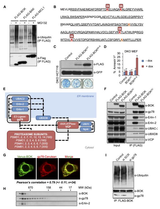
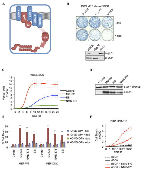
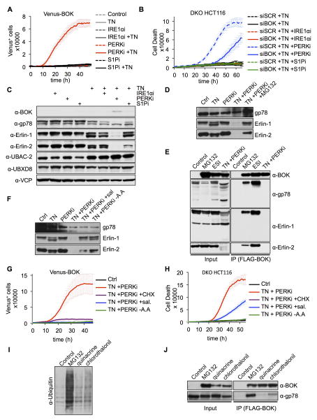
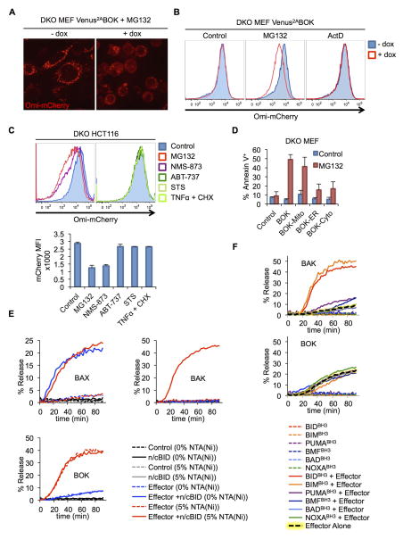
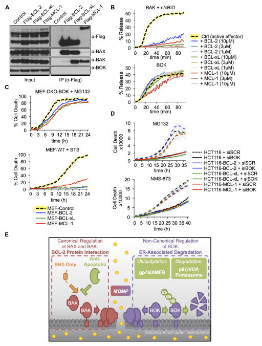
The mitochondrial pathway of apoptosis is initiated by mitochondrial outer membrane permeabilization (MOMP). The BCL-2 family effectors BAX and BAK are thought to be absolutely required for this process. Here, we report that BCL-2 ovarian killer (BOK) is a bona fide yet unconventional effector of MOMP that can trigger apoptosis in the absence of both BAX and BAK. However, unlike the canonical effectors, BOK appears to be constitutively active and unresponsive to antagonistic effects of the antiapoptotic BCL-2 proteins. Rather, BOK is controlled at the level of protein stability by components of the endoplasmic reticulum (ER)-associated degradation pathway. BOK is ubiquitylated by the AMFR/gp78 E3 ubiquitin ligase complex and targeted for proteasomal degradation in a VCP/p97-dependent manner, which allows survival of the cell. When proteasome function, VCP, or gp78 activity is compromised, BOK is stabilized to induce MOMP and apoptosis independently of other BCL-2 proteins.


Conflict of interest statement

The authors declare no conflict of interest.

Figures

Figure 1
Figure 1. BOK triggers apoptosis in response to proteasome inhibition
A. IncuCyte quantification of Venus-BOK in WT MEFs treated with dox, MG132 (10 μM), bortezomib (1 μM), carfilzomib (1 μM), actinomycin D (1 μM), etoposide (100 μM), UV (10 mJ/cm2), staurosporine (1 μM), thapsigargin (1 μM) or TN (1 μg/mL), and Q-VD-POh (40 μM). B. Immunoblot of FLAG-tagged BCL-2 family proteins transfected in 293T cells in the presence of 40 μM Q-VD-POh. C. Immunoblot of Venus and BOK. WT MEFs expressing Venus2ABOK were treated as in (A). D. IncuCyte quantification of SYTOX Green–stained WT MEFs expressing Venus2ABOK treated as in (A), in the absence of Q-VD-POh. E. Clonogenic survival of cancer cells treated with scrambled (SCR) or BOK-targeting siRNA and 10 μM MG132 for 30 h. F. Flow cytometry quantification of Annexin V–stained HCT116, T98G, and A172 cells transfected with siRNA and treated with 10 μM MG132 for 30 h (mean ± SD of 3 independent experiments). G. IncuCyte quantification of SYTOX Green–stained HCT116 cells treated with siRNA and MG132 (10 μM), ABT-737 (30 μM), staurosporine (2 μM) or a combination of TNFα (50 ng/mL) and cycloheximide (CHX, 2 μg/mL). IncuCyte data are represented as mean of triplicate samples ± SD and representative of 3 independent experiments (see also Figure S1).
Figure 2
Figure 2. BOK triggers apoptosis independently of BAX and BAK
A. IncuCyte quantification of SYTOX Green-stained bax−/−bak−/− MEFs expressing Venus2ABOK and treated as in 1(A), in the absence of Q-VD-POh. B. Confocal microscopy images of bax−/−bak−/− MEFs expressing Venus or Venus2ABOK treated with dox, Annexin V Alexa Fluor® 647, and 10 μM MG132 for 3 h. C. IncuCyte quantification of SYTOX Green–stained WT or bax−/−bak−/− HCT116 cells treated with scrambled (SCR) or BOK-targeting siRNA and TNFα (50 ng/mL) plus cycloheximide (CHX, 2 μg/mL) or MG132 (10 μM). D. Flank tumor volume of CD-1 nude mice injected with bax−/−bak−/− MEFs expressing Venus (left flank) or Venus2ABOK (right flank) and treated with bortezomib at days 4 and 8 (right panel). E. Summary of the high-throughput screening of 4282 biologically active compounds for caspase activation in bax−/−bak−/− MEFs expressing Venus2ABOK. IncuCyte data are represented as mean of triplicate samples ± SD and representative of 3 independent experiments (see also Figure S2 and Movies S1 and S2).
Figure 3
Figure 3. BOK stability is regulated by the ERAD pathway
A. Ubiquitylation profile of Flag-tagged BOK, BAK, or MCL-1 immunoprecipitated from transiently transfected HCT116 cells treated with 40 μM Q-VD-POh ±10 μM MG132. B. Ubiquitylation sites in murine BOK identified by MS (peptide coverage underlined). C. Clonogenic survival of bax−/−bak−/− HCT116 cells transiently transfected with an empty pMX-IRES-GFP or expressing FLAG-BOKWT or FLAG-BOK6K-R and sorted for equivalent GFP intensity (lower panel). Upper panel: immunoblot of FLAG and GFP. D. Flow cytometry quantification of Annexin V–stained bax−/−bak−/− MEFs expressing Venus2ABOKWT or Venus2ABOK6K-R and treated with dox for 24 h (mean ± SD of 3 independent experiments) E. BOK interacting network as identified by tandem affinity purification and MS analysis. F. Immunoprecipitation of Flag-BOKFL and Flag-BOKΔC from lysates of bax−/−bak−/− MEFs treated with dox, MG132 (10 μM), and Q-VD-OPh (40 μM). G. Confocal microscopy images of HeLa cells cotransfected with Venus-BOK and gp78-Cerulean in presence of MG132 (10 μM) and Q-VD-OPh. Pearson correlation coefficient ± SD is indicated. H. Size exclusion chromatography of cell lysates from MEFs expressing Venus2ABOK and treated with dox, MG132 (10 μM), and Q-VD-OPh (40 μM). I. Ubiquitylation profile of Flag-tagged BOK immunoprecipitated from bax−/−bak−/− MEFs treated with scrambled (SCR) or gp78 targeting siRNA and dox, MG132 (10 μM), and Q-VD-OPh (40 μM) (see also Figure S3).
Figure 4
Figure 4. ERAD disruption triggers BOK-induced apoptosis
A. Schematic representation of ERAD components regulating BOK degradation. B. Clonogenic survival of bax−/−bak−/− MEFs expressing Venus2ABOK transfected with scrambled (SCR), gp78-, or VCP-targeting siRNA and treated with dox for 24 h. C. IncuCyte quantification of Venus-BOK in bax−/−bak−/− MEFs treated with dox and MG132 (10 μM), ESI (1μM), and NMS-873 (10μM) in the presence of 40 μMQ-VD-OPh. D. Immunoblot of Venus and BOK. bax−/−bak−/− MEFs expressing Venus2ABOK were treated as in (C). E. Flow cytometry quantification of Annexin V–stained WT and bax−/−bak−/− MEFs expressing Venus2ABOK and treated with dox and MG132 (10 μM), ESI (1 μM) or NMS-873 (10 μM) for 8 h (mean ± SD of 3 independent experiments). F. IncuCyte quantification of SYTOX Green–stained bax−/−bak−/− HCT116 cells treated with scrambled (SCR) or BOK-targeting siRNA and 10 μM NMS-873. IncuCyte data represented as mean of triplicate samples ± SD and representative of 3 independent experiments (see also Figure S4).
Figure 5
Figure 5. Inhibition of PERK during ER stress induces ERAD disruption and BOK-induced apoptosis
A. IncuCyte quantification of Venus-BOK in bax−/−bak−/− MEFs treated with dox, Q-VD-OPh (40 μM), TN (1 μg/mL) and 4μ8C (IRE1αi, 50 μM), GSK2656157 (PERKi, 500 nM), or PF429242 (S1Pi, 10 μM). B. IncuCyte quantification of SYTOX Green– stained bax−/−bak−/− HCT116 cells transfected with scrambled (SCR) or BOK targeting siRNA and treated as in (A), in the absence of QV-VD-OPh. C, D. Immunoblot of lysate from bax−/−bak−/− MEFs expressing Venus2ABOK and treated as in (A). E. Immunoprecipitation of FLAG-BOK from lysate of bax−/−bak−/− MEFs treated with dox, Q-VD-OPh (40 μM), and MG132 (10 μM), ESI (1 μM) or TN (1 μg/mL) plus GSK2656157 (PERKi, 500 nM). NB: Immunoprecipitated samples were normalized for equivalent BOK loading. F. Immunoblot of bax−/−bak−/− MEF lysates treated with Q-VD-OPh, TN (1 μg/mL), GSK2656157 (PERKi, 500 nM), and salubrinal (100 μM) in complete DMEM or HBSS medium (-A.A). G. IncuCyte quantification of Venus-BOK in bax−/−bak−/− MEFs treated as in (F). H. IncuCyte quantification of SYTOX Green–stained bax−/−bak−/− HCT116 cells treated as in (F), in the absence of Q-VD-OPh. I. Global protein ubiquitylation of lysates from bax−/−bak−/− MEFs expressing FLAG-BOK and treated with dox, Q-VD-OPh (40 μM), and MG132 (10 μM), quinacrine (20 μM), or chlorothalonil (2 μM). J. Immunoprecipitation of FLAG-BOK from lysates of bax−/−bak−/− MEFs treated as in (I). NB: Immunoprecipitated samples were normalized for equivalent BOK loading. IncuCyte data are represented as mean of triplicate samples ± SD and representative of 3 independent experiments (see also Figure S5).
Figure 6
Figure 6. BOK is a constitutively active BCL-2 family effector
A. Confocal microscopy images of bax−/−bak−/− MEFs expressing Omi-mCherry and Venus2ABOK treated with dox, Q-VD-OPh (40 μM), and MG132 (10 μM) for 3 h. B. MOMP was assessed through flow cytometry quantification of loss of Omi-mCherry from digitonin-permeabilized bax−/−bak−/− MEFs treated as in (A). C. bax−/−bak−/− HCT116 expressing Omi-mCherry were processed as in (B) (left histogram: treatment with MG132 (10 μM), NMS-873 (10 μM); right histogram: treatment with ABT-737 (30 μM), staurosporine (STS, 2 μM), TNFα (50 ng/mL) plus cycloheximide (CHX, 2 μg/mL). D. Flow cytometry quantification of Annexin V–stained bax−/−bak−/− MEFs expressing BOK targeted constructs and treated with dox and 10 μM MG132 for 8 h (mean ± SD of 3 independent experiments). E. Permeabilization of LUV containing 0% or 5% DGS-NTA(Ni), upon incubation with BAXFL (100 nM), BAKΔC-8xHIS (1 μM) or BOKΔC-8xHIS (1 μM) and cleaved BID (n/cBID, 20 nM). F. NTA(Ni)-LUV permeabilization upon incubation with BH3 peptides (5 μM) and BAKΔC-8xHIS (1 μM) or BOKΔC-8xHIS (50 nM). LUV data are represented as mean of triplicate samples and representative of 3 independent experiments (see also Figures S6 and S7 and Movies S3 and S4).
Figure 7
Figure 7. BOK is not inhibited by antiapoptotic BCL-2 proteins
A. Immunoprecipitation of FLAG-tagged BCL-2, BCL-xL, or MCL-1 from lysates of WT MEFs expressing BOK and treated with dox, MG132 (10 μM), and Q-VD-OPh (40 μM). B. NTA(Ni)-LUV upon incubation with n/cBID (20 nM) + BAKΔC-8xHIS (1 μM) or BOKΔC-8xHIS (1 μM) in combination with BCL-2 ΔC-8xHIS, BCL-xL ΔC-8xHIS, or MCL-1 ΔC-8xHIS. LUV data are represented as mean of triplicate samples ± SD and representative of 3 independent experiments. C. IncuCyte quantification of SYTOX Green–stained WT or bax−/−bak−/− MEFs expressing Venus2ABOK and overexpressing BCL-2, BCL-xL, or MCL-1 treated with the MG132 (10 μM) or staurosporine (1 μM). D. IncuCyte quantification of SYTOX Green–stained DKO HCT116 cells overexpressing BCL-2, BCL-xL, or MCL-1, treated with scrambled (SCR) or BOK-targeting siRNA and 10 μM MG132 (upper panel) or 10 μM NMS-873 (lower panel). E. Model of BOK-induced MOMP regulation by the ERAD pathway, independently of the BCL-2 protein family. IncuCyte data are represented as mean of triplicate samples ± SD representative of 3 independent experiments (see also Figure S7).

Comment in

References

    1. Antonsson B, Montessuit S, Lauper S, Eskes R, Martinou JC. Bax oligomerization is required for channel-forming activity in liposomes and to trigger cytochrome c release from mitochondria. Biochem J. 2000;345(Pt 2):271–278. - PMC - PubMed
    1. Asciolla JJ, Renault TT, Chipuk JE. Examining BCL-2 family function with large unilamellar vesicles. J Vis Exp. 2012;68:e4291. - PMC - PubMed
    1. Atkins C, Liu Q, Minthorn E, Zhang SY, Figueroa DJ, Moss K, Stanley TB, Sanders B, Goetz A, Gaul N, et al. Characterization of a novel PERK kinase inhibitor with antitumor and antiangiogenic activity. Cancer Res. 2013;73:1993–2002. - PubMed
    1. Back SH, Scheuner D, Han J, Song B, Ribick M, Wang J, Gildersleeve RD, Pennathur S, Kaufman RJ. Translation attenuation through eIF2alpha phosphorylation prevents oxidative stress and maintains the differentiated state in beta cells. Cell Metab. 2009;10:13–26. - PMC - PubMed
    1. Beroukhim R, Mermel CH, Porter D, Wei G, Raychaudhuri S, Donovan J, Barretina J, Boehm JS, Dobson J, Urashima M, et al. The landscape of somatic copy-number alteration across human cancers. Nature. 2010;463:899–905. - PMC - PubMed

Publication types

MeSH terms

Substances