Review
. 2016 May:64:196-214.
doi: 10.1016/j.neubiorev.2016.02.020. Epub 2016 Mar 3.

Social dysfunction after pediatric traumatic brain injury: A translational perspective

Affiliations
Review

Social dysfunction after pediatric traumatic brain injury: A translational perspective

Nicholas P Ryan et al. Neurosci Biobehav Rev. 2016 May.

Abstract

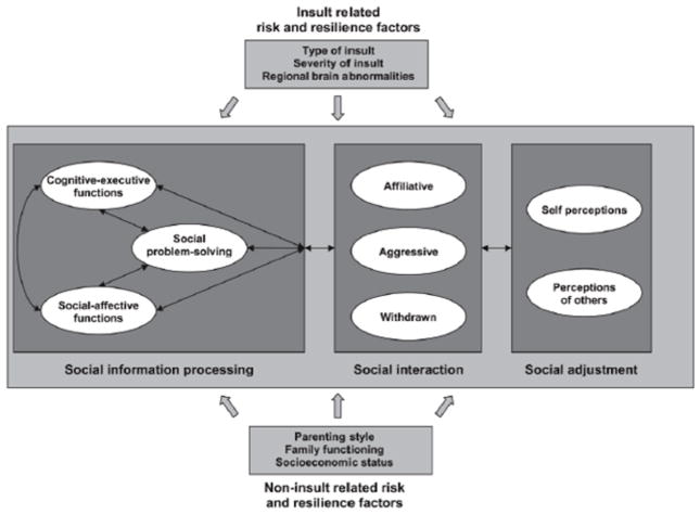
Social dysfunction is common after traumatic brain injury (TBI), contributing to reduced quality of life for survivors. Factors which influence the development or persistence of social deficits after injury remain poorly understood, particularly in the context of ongoing brain maturation during childhood and adolescence. Aberrant social interactions have recently been modeled in adult and juvenile rodents after experimental TBI, providing an opportunity to gain new insights into the underlying neurobiology of these behaviors. Here, we review our current understanding of social dysfunction in both humans and rodent models of TBI, with a focus on brain injuries acquired during early development. Modulators of social outcomes are discussed, including injury-related and environmental risk and resilience factors. Disruption of social brain network connectivity and aberrant neuroendocrine function are identified as potential mechanisms of social impairments after pediatric TBI. Throughout, we highlight the overlap and disparities between outcome measures and findings from clinical and experimental approaches, and explore the translational potential of future research to prevent or ameliorate social dysfunction after childhood TBI.

Keywords: Behavior; Brain; Children; Communication; Humans; Pediatric; Rodents; Social brain network; Social competency; Social interactions; Traumatic brain injury.


Figures

Figure 1
Figure 1
Heuristic model of social competence. Reproduced with Yeates et al. (2007), with permission from the American Psychological Association.
Figure 2
Figure 2
Tripartite model of Theory of Mind. Reproduced from Dennis et al. (2013), with permission from Elsevier.
Figure 3
Figure 3
Assays for social behavior in rodents. A range of experimental paradigms can be used to evaluate social behaviors between rodents, including the evaluation of social interactions in a neutral environment (a) or home cage of the test animal (b). The three-chamber apparatus provides a measure of sociability (c) and subsequent evaluation of social recognition memory (d). Social behaviors can alternatively be examined within a group housing context (e). Social communication may be quantified by the deposition of urinary scent marks in response to a conspecific (f). Adapted from Sandi (2015).
Figure 4
Figure 4
The social brain network in humans and rodents. Key brain regions involved in the social brain network in humans and rodents. PFC, prefrontal cortex; ACC, anterior cingulate cortex; cc, corpus callosum; Th, thalamus; Am, amygdala; NAc, nucleus accumbens; Hpc, hippocampus; Hyp, hypothalamus; PAG, periaqueductal gray.
Figure 5
Figure 5
Distribution of brain lesions in the social brain network after pediatric TBI. Probability distribution of brain lesions detected using SWI in the left lateral (A), left medial (B), right lateral (C) and right medial (D) hemispheres. Lesion distributions were created by aligning the individual T1 images to the Montreal Neurological Institute template using the non-linear normalization procedure in Statistical Parametric Mapping 8 (SPM8). The lesion maps were normalized using the same transformations. The aligned lesion masks were averaged to produce a single image illustrating the distribution of lesions in the study population. Hotter colors indicate the co-location of lesions in multiple subjects. Lesions were most prominent in frontal regions [frontal only = 15 patients, frontal + extrafrontal only = 6, frontal + other regions (CC (corpus callosum) = 1, deep gray + CC = 1, cerebellum = 1, cerebellum + CC = 1)], followed by extrafrontal regions only (N = 6). A small number of patients (4) had lesions in several areas (frontal + extrafrontal + cerebellum = 2, frontal + extrafrontal + deep gray = 1, frontal + extrafrontal + CC = 1). Very few patients had lesions solely in the CC (1), cerebellum (1) or deep gray (0) regions. Reproduced from Beauchamp et al. (2013).

References

    1. Agoston DV. Bench-to-Bedside and Bedside Back to the Bench; Seeking a Better Understanding of the Acute Pathophysiological Process in Severe Traumatic Brain Injury. Front Neurol. 2015;6:47. - PMC - PubMed
    1. Amaral DG, Bauman MD, Schumann CM. The amygdala and autism: implications from non-human primate studies. Genes Brain Behav. 2003;2:295–302. - PubMed
    1. Ameis SH, Catani M. Altered white matter connectivity as a neural substrate for social impairment in Autism Spectrum Disorder. Cortex. 2015;62:158–181. - PubMed
    1. Anderson V, Beauchamp MH, Yeates KO, Crossley L, Hearps SJC, Catroppa C. Social Competence at 6 Months Following Childhood Traumatic Brain Injury. J Int Neuropsychol Soc. 2013;19:539–550. - PubMed
    1. Anderson V, Catroppa C, Morse S, Haritou F, Rosenfeld J. Functional plasticity or vulnerability after early brain injury? Pediatrics. 2005;116:1374–1382. - PubMed

Publication types