. 2016 Mar 7:5:e13046.
doi: 10.7554/eLife.13046.

Cryo-EM single particle analysis with the Volta phase plate

Affiliations

Cryo-EM single particle analysis with the Volta phase plate

Radostin Danev et al. Elife. .

Abstract

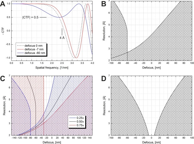
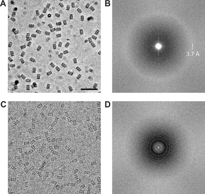
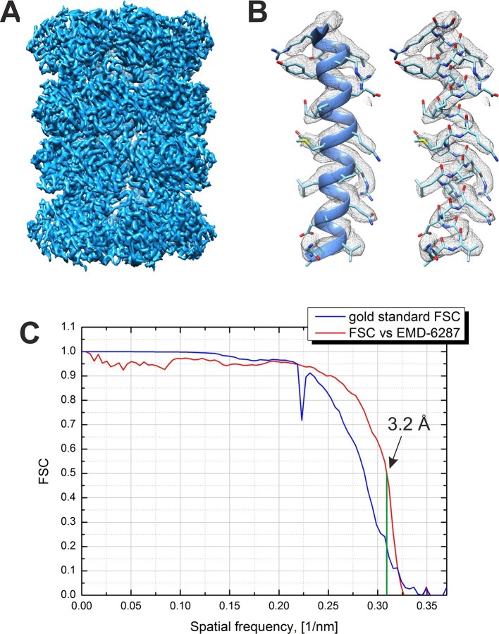
We present a method for in-focus data acquisition with a phase plate that enables near-atomic resolution single particle reconstructions. Accurate focusing is the determining factor for obtaining high quality data. A double-area focusing strategy was implemented in order to achieve the required precision. With this approach we obtained a 3.2 Å resolution reconstruction of the Thermoplasma acidophilum 20S proteasome. The phase plate matches or slightly exceeds the performance of the conventional defocus approach. Spherical aberration becomes a limiting factor for achieving resolutions below 3 Å with in-focus phase plate images. The phase plate could enable single particle analysis of challenging samples in terms of small size, heterogeneity and flexibility that are difficult to solve by the conventional defocus approach.

Keywords: biophysics; cryo-EM; none; phase plate; single particle analysis; structural biology.


Conflict of interest statement

RD: Co-inventor in US patent US9129774 B2 "Method of using a phase plate in a transmission electron microscope".

WB: Scientific Advisory Board of FEI Company.

Figures

Figure 1.
Figure 1.. Volta phase plate CTF examples and allowed defocus ranges versus resolution.
(A) Illustration of CTFs at defocus values that limit the resolution to 4 Å according to a |CTF|=0.5 criterion. (B) Defocus limits versus resolution according to the |CTF|=0.5 criterion for a π/2 phase plate and 2.7 mm spherical aberration. The shaded areas are 'prohibited' in a sense that for those defocus values the CTF amplitude drops below 0.5 at a resolution lower than the value on the y-axis. (C) Same as (B) but for three different phase shift values. (D) Same as (B) but for a Cs-corrected microscope (0 mm spherical aberration). DOI: http://dx.doi.org/10.7554/eLife.13046.003
Figure 2.
Figure 2.. Data acquisition schemes that enable precise focusing superposed on a holey support film.
(A) Two focusing areas, F1 and F2, on opposite sides of the data acquisition position D are used to perform a linear interpolation of the measured defocus. (B) Multiple focusing areas F1 to F4 around a support film hole can be used to interpolate the defocus and acquire multiple data images (D1 to D4) per hole. DOI: http://dx.doi.org/10.7554/eLife.13046.004
Figure 3.
Figure 3.. Measured phase shift of the VPP as a function of the image number/total dose on the phase plate.
The measurement was performed at two consecutive positions on the phase plate. DOI: http://dx.doi.org/10.7554/eLife.13046.005
Figure 4.
Figure 4.. Representative images from 20S proteasome datasets acquired with and without a phase plate.
(A) In-focus image acquired with the Volta phase plate. (B) Power spectrum of the image in (A). The presence of the amorphous ice ring at 3.7 Å indicates that there is good information transfer to at least that spatial frequency. (C) Conventional defocus image at −1.6 μm defocus. (D) Power spectrum of the image in (C) showing CTF Thon rings. Scale bar: 50 nm. DOI: http://dx.doi.org/10.7554/eLife.13046.006
Figure 5.
Figure 5.. Result from the 3D reconstruction of 20S proteasome from an in-focus dataset acquired with the Volta phase plate.
(A) An isosurface representation of the density map. (B) A ribbon and a stick representations of an α-helical segment from the β subunit docked into the density map demonstrating the presence of sidechain densities. (C) Fourier shell correlation (FSC) curves from the Relion software’s internal 'gold standard' and from a comparison with an external 2.8 Å density map (EMD-6287). Both criteria indicate a resolution of 3.2 Å at a 0.143 level for the 'gold standard' FSC and at a 0.5 level for the external map. The Nyquist frequency is at 2.7 Å (0.37 1/nm). DOI: http://dx.doi.org/10.7554/eLife.13046.007
Figure 6.
Figure 6.. Resolution versus number of particles and B-factor estimations.
(A) Resolution versus number of particles for several datasets. The resolutions were calculated based on the 0.5 FSC level versus an external 2.8 Å density map (EMD-6287). For two of the datasets (red and blue lines) the number of particles was varied artificially by using random subsets of particles. (B) Logarithm of the number of particles versus the squared reciprocal resolution plots of the two main datasets in (A). The legend contains B-factors estimated by linear fits of two data regions. DOI: http://dx.doi.org/10.7554/eLife.13046.008

Comment in

  • Protein complexes in focus.
    Glaeser RM. Glaeser RM. Elife. 2016 Apr 28;5:e16156. doi: 10.7554/eLife.16156. Elife. 2016. PMID: 27124202 Free PMC article.

Similar articles

Cited by

References

    1. Asano S, Fukuda Y, Beck F, Aufderheide A, Forster F, Danev R, Baumeister W. A molecular census of 26S proteasomes in intact neurons. Science. 2015;347:439–442. doi: 10.1126/science.1261197. - DOI - PubMed
    1. Bartesaghi A, Merk A, Banerjee S, Matthies D, Wu X, Milne JLS, Subramaniam S. 2.2 Å resolution cryo-EM structure of beta-galactosidase in complex with a cell-permeant inhibitor. Science. 2015;348:1147–1151. doi: 10.1126/science.aab1576. - DOI - PMC - PubMed
    1. Campbell MG, Veesler D, Cheng A, Potter CS, Carragher B. 2.8 Å resolution reconstruction of the Thermoplasma acidophilum 20S proteasome using cryo-electron microscopy. eLife. 2015;4:e13046. doi: 10.7554/eLife.06380. - DOI - PMC - PubMed
    1. Chang W-H, Chiu MT-K, Chen C-Y, Yen C-F, Lin Y-C, Weng Y-P, Chang J-C, Wu Y-M, Cheng H, Fu J, Tu I-P. Zernike phase plate cryoelectron microscopy facilitates single particle analysis of unstained asymmetric protein complexes. Structure. 2010;18:17–27. doi: 10.1016/j.str.2009.12.001. - DOI - PubMed
    1. Chen JZ, Grigorieff N. SIGNATURE: A single-particle selection system for molecular electron microscopy. Journal of Structural Biology. 2007;157:168–173. doi: 10.1016/j.jsb.2006.06.001. - DOI - PubMed

Publication types

MeSH terms

Substances