. 2016 Mar 7;11(3):e0150878.
doi: 10.1371/journal.pone.0150878. eCollection 2016.

Polo-Like Kinase 3 Appears Dispensable for Normal Retinal Development Despite Robust Embryonic Expression

Affiliations

Polo-Like Kinase 3 Appears Dispensable for Normal Retinal Development Despite Robust Embryonic Expression

Jillian J Goetz et al. PLoS One. .

Abstract

During retinogenesis seven different cell types are generated in distinct yet overlapping timepoints from a population of retinal progenitor cells. Previously, we performed single cell transcriptome analyses of retinal progenitor cells to identify candidate genes that may play roles in the generation of early-born retinal neurons. Based on its expression pattern in subsets of early retinal cells, polo-like kinase 3 (Plk3) was identified as one such candidate gene. Further characterization of Plk3 expression by in situ hybridization revealed that this gene is expressed as cells exit the cell cycle. We obtained a Plk3 deficient mouse and investigated changes in the retina's morphology and transcriptome through immunohistochemistry, in situ hybridization and gene expression profiling. These experiments have been performed initially on adult mice and subsequently extended throughout retinal development. Although morphological studies revealed no consistent changes in retinogenesis upon Plk3 loss, microarray profiling revealed potential candidate genes altered in Plk3-KO mice. Further studies will be necessary to understand the connection between these changes in gene expression and the loss of a protein kinase such as Plk3.


Conflict of interest statement

Competing Interests: The authors have declared that no competing interests exist.

Figures

Fig 1
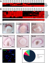
Fig 1. Survey of Plk3 expression in the developing mouse retina.
(A) A heatmap showing the expression of Plk family members in Math5-positive cells. The four E12 cells at the left are Math5-negative, but were previously identified as G2/M progenitor cells [2]. These cells are shown as a comparison. In the heatmap, the genes (in rows) expressed in isolated single retinal progenitor cells (in columns) at various stages of development from embryonic day (E)12.5 to E16.5 are shown. Higher levels of microarray signal of a given gene correspond to higher expression levels in a particular single cell and are indicated by the different shades of red (see the scale below [B]), while the absence of expression is indicated with a black square. (B) A heatmap showing G2/M cell cycle marker expression in Math5 and Plk3 expressing single cells. As in (A) the log transformed signal intensities have been scaled according to the intensity of red color, with black indicating the absence of expression signal. The eleven single cells shown on the left are included for comparison and are Plk3-negative cells that were previously identified as in the G2/M phase of the cell cycle [2]. (C) In situ hybridization of Plk3 expression in the embryonic mouse retina. (D) In situ hybridization showing the expression of Tcfap2b, Tcfap2d, and Nhlh2, markers of developing amacrine interneurons, at E14.5. (E) Double fluorescent in situ hybridization of Math5 (green) and Plk3 (red) at E14.5. The results are quantified in (F). All scale bars represent 100 μm.
Fig 2
Fig 2. Morphological characterization of adult Plk3-KO retinas.
Populations of adult (>P35) retinal cells were identified using antibodies to neuron-specific markers. DAPI, in blue, marks nuclei. Rod photoreceptors (anti-Rhodopsin [Rho4d2], A,A’), bipolar interneurons (anti-Chx10, B,B’; anti-PKC-α, C,C’), Müller glia (anti-Glutamine synthetase [GS] D,D’), amacrine interneurons (anti-Pax6, E,E’; anti-Chat, F,F’), retinal ganglion cells (anti-Brn3b, G,G’), and horizontal, amacrine and ganglion cells (anti-Calretinin [Calr], anti-Calbindin28k [Calb28k], H,H’) are shown. Scale bars represent 100 μm.
Fig 3
Fig 3. Assessment of cell numbers by flat mount antibody staining.
To more quantitatively assess any gains/losses in cell number in the Plk3-KO retina, immunohistochemistry was performed on flat mounted retinas from adult (>P35) wildtype and Plk3-KO littermates. Confocal scans were performed on four different quadrants from each retina and representative quadrant-matched images are shown. DAPI, in blue, marks nuclei. Horizontal cells (anti-Hnf6, A,A’; anti-Calbindin28k [Calb28k], B,B’), amacrine interneurons (anti-Ap2a, C,C’), a combination of amacrine and ganglion cells (anti-Calretinin [Calr], D,D’) and retinal ganglion cells (anti-Brn3a, E,E’; anti-Opn4, F,F’) are shown. Scale bars represent 100 μm.
Fig 4
Fig 4. Morphological characterization of postnatal developing Plk3-KO retinas.
Populations of retinal cells in postnatal day (P)7 Plk3-KO retinas were compared to WT littermates using cell type-specific markers. Specifically, populations of rod photoreceptors (anti-Rhodopsin [Rho4d2], A,A’), bipolar cells (anti-Chx10, B,B’), retinal ganglion cells (anti-Brn3b, C,C’), bipolar cells, amacrine cells and ganglion cells (anti-Isl1, D, D’), amacrine, horizontal and ganglion cells (anti-Pax6, E,E’; anti-Calretinin [Calr], anti-Calbindin28k [Calb28k], F,F’) were examined. Scale bars indicate 100 μm.
Fig 5
Fig 5. Survey of retinal cell types in P4 Plk3-KO retinas.
Populations of retinal cells in P4 Plk3-KO retinas were compared to WT littermates using cell type-specific markers for photoreceptors (anti-Recoverin, A,A’), bipolar cells (anti-Chx10, B,B’), retinal ganglion cells (anti-Brn3b, C,C’), ganglion and amacrine cells (anti-Isl1, D,D’; anti-Pax6, E,E’) and amacrine, horizontal and ganglion cells (anti-Calretinin [Calr], anti-Calbindin28k [Calb28k], F,F’). Scale bars indicate 100 μm.
Fig 6
Fig 6. Survey of retinal cell types in P14 Plk3-KO retinas.
Populations of retinal cells in P14 Plk3-KO retinas were compared to WT littermates using cell type-specific markers for rod photoreceptors (anti-Rhodopsin [Rho4d2], A,A’), bipolar cells (anti-Chx10, B,B’), retinal ganglion cells (anti-Brn3b, C,C’), ganglion and amacrine cells (anti-Isl1, D,D’; anti-Pax6, E,E’) and amacrine, horizontal and ganglion cells (anti-Calretinin [Calr], anti-Calbindin28k [Calb28k], F,F’). Scale bars indicate 100 μm.
Fig 7
Fig 7. qPCR based examination of gene expression in Plk3-KO retinas.
Retinas of Plk3-KO and WT littermates were isolated and hybridized to Affymetrix microarrays at various timepoints (n = 3 for each of four timepoints). Genes with significant differential expression at multiple timepoints were confirmed using qPCR at adult timepoints. (A) An examination of Plk3 expression in the Plk3 deficient mouse. An amplicon at the 3’ end of the gene showed upregulation (p<0.001), while one at the 5’ end of Plk3 was significantly downregulated (p<0.05). (B) Tac1 showed decreased expression (p<0.05), whereas retinitis pigmentosa 1 (Rp1) displayed increased expression (p<0.05).
Fig 8
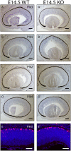
Fig 8. Examination of Plk family member expression patterns and cell cycle at E14.5.
The expression patterns of Plk3 (A, A’), Plk1 (B,B’), Plk2 (C,C’), and Plk4 (D,D’) in WT and Plk3-KO retinas were determined using in situ hybridization at E14.5. E14.5 developing WT and Plk3-KO retinas were also stained using anti-phospho-histone H3 (PH3) to mark mitotic cells (E,E’).

Similar articles

Cited by

References

    1. Goetz JJ, Farris C, Chowdhury R, Trimarchi JM. Making of a retinal cell: insights into retinal cell-fate determination. International review of cell and molecular biology. 2014;308:273–321. Epub 2014/01/15. 10.1016/B978-0-12-800097-7.00007-5 . - DOI - PubMed
    1. Trimarchi JM, Stadler MB, Cepko CL. Individual retinal progenitor cells display extensive heterogeneity of gene expression. PLoS ONE. 2008;3(2):e1588 Epub 2008/02/14. 10.1371/journal.pone.0001588 - DOI - PMC - PubMed
    1. Masland RH. The fundamental plan of the retina. Nat Neurosci. 2001;4(9):877–86. Epub 2001/08/31. 10.1038/nn0901-877 . - DOI - PubMed
    1. Young RW. Cell differentiation in the retina of the mouse. Anat Rec. 1985;212(2):199–205. . - PubMed
    1. Young RW. Cell proliferation during postnatal development of the retina in the mouse. Brain Res. 1985;353(2):229–39. . - PubMed

Publication types

MeSH terms

Substances