Review
. 2016 Mar 2;17(3):273.
doi: 10.3390/ijms17030273.

The Immunology of Neuromyelitis Optica-Current Knowledge, Clinical Implications, Controversies and Future Perspectives

Affiliations
Review

The Immunology of Neuromyelitis Optica-Current Knowledge, Clinical Implications, Controversies and Future Perspectives

Michalina Jasiak-Zatonska et al. Int J Mol Sci. .

Abstract

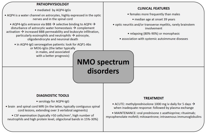
Neuromyelitis optica (NMO) is an autoimmune, demyelinating disorder of the central nervous system (CNS) with typical clinical manifestations of optic neuritis and acute transverse myelitis attacks. Previously believed to be a variant of multiple sclerosis (MS), it is now considered an independent disorder which needs to be differentiated from MS. The discovery of autoantibodies against aquaporin-4 (AQP4-IgGs) changed our understanding of NMO immunopathogenesis and revolutionized the diagnostic process. AQP4-IgG is currently regarded as a specific biomarker of NMO and NMO spectrum disorders (NMOsd) and a key factor in its pathogenesis. Nevertheless, AQP4-IgG seronegativity in 10%-25% of NMO patients suggests that there are several other factors involved in NMO immunopathogenesis, i.e., autoantibodies against aquaporin-1 (AQP1-Abs) and antibodies against myelin oligodendrocyte glycoprotein (MOG-IgGs). This manuscript reviews current knowledge about NMO immunopathogenesis, pointing out the controversial issues and showing potential directions for future research. Further efforts should be made to broaden our knowledge of NMO immunology which could have important implications for clinical practice, including the use of potential novel biomarkers to facilitate an early and accurate diagnosis, and modern treatment strategies improving long-term outcome of NMO patients.

Keywords: aquaporin-1 antibody (AQP1-Ab); aquaporin-4 immunoglobulin G (AQP4-IgG); immunopathogenesis; myelin oligodendrocyte glycoprotein immunoglobulin G (MOG-IgG); neuroimmunology; neuromyelitis optica (NMO); neuromyelitis optica spectrum disorder (NMOsd).


Figures

Figure 1
Figure 1
Diagnostic criteria for neuromyelitis spectrum according to International Panel for Neuromyelitis Optica (NMO) Diagnosis (2015) [8]. NMOsd: NMO spectrum disorders; AQP4-IgG: aquaporin-4 immunoglobulin G; MRI: magnetic resonance imaging.
Figure 2
Figure 2
Neuroradiology of neuromyelitis optica spectrum disorder (NMOsd): (a,b) magnetic resonance imaging (MRI) visualisation of cervical spinal cord lesions in a 51-year old female with AQP4-IgG (+) NMOsd; (a) longitudinally extensive, central, extending to brainstem T2-hyperintense lesion; (b) corresponding T1-hypointensities, representing focal spinal cord atrophy; (c,d) thoracic spinal cord and brain MRI lesions in a 20-year old AQP4-IgG (+) female with NMOsd; (c) nearly complete central longitudinal thoracic spinal cord NMO involvement; (d) brain lesions on FLAIR (fluid-attenuated inversion recovery) images that do not fulfill multiple sclerosis diagnostic criteria. Images from the collection of Department of Neurology, Poznan University of Medical Sciences.
Figure 3
Figure 3
Immunopathogenesis of neuromyelitis optica: (1) primary immunizing event of unknown provenance; (2) T lymphocytes take part in breaking the tolerance and in recruitment of other leukocytes; (3) plasmablasts produce AQP4-IgGs, which enter the central nervous system (CNS) through (4) endothelial transcytosis or at areas of increased blood-brain barrier permeability and then bind selectively to aquaporin-4 (AQP4); (5) complement activation, leading to complement-dependent cytotoxicity and subsequent astrocyte death; (6) neutrophil and eosinophil rise in the periphery and their subsequent CNS infiltration (7) neutrophil and eosinophil infiltration (8) secondary demyelination caused by, among others, myelin phagocytosis, and bystander injury by pro-inflammatory factors. APC: antigen presenting cell; MAC: the membrane attack complex.
Figure 4
Figure 4
Immunofluorescence staining for aquaporin-4 in a cell-based assay: (a) negative stain; (b) positive stain. Images from the collection of Division of Neurochemistry and Neuropathology, Department of Neurology, Poznan University of Medical Sciences.
Figure 5
Figure 5
Basic summary of the current knowledge on NMO spectrum disorders. Abbreviations: AQP4—aquaporin 4; BBB—blood-brain barrier; IgG—immunoglobulin G; MRI—magnetic resonance imaging; CSF—cerebrospinal fluid.

References

    1. Wingerchuk D.M., Hogancamp W.F., O’Brien P.C., Weinshenker B.G. The clinical course of neuromyelitis optica (Devic’s syndrome) Neurology. 1999;53:1107. doi: 10.1212/WNL.53.5.1107. - DOI - PubMed
    1. Wingerchuk D.M., Lennon V.A., Lucchinetti C.F., Pittock S.J., Weinshenker B.G. The spectrum of neuromyelitis optica. Lancet Neurol. 2007;6:805–815. doi: 10.1016/S1474-4422(07)70216-8. - DOI - PubMed
    1. Wingerchuk D.M., Lennon V.A., Pittock S.J., Lucchinetti C.F., Weinshenker B.G. Revised diagnostic criteria for neuromyelitis optica. Neurology. 2006;66:1485–1489. doi: 10.1212/01.wnl.0000216139.44259.74. - DOI - PubMed
    1. Jarius S., Wildemann B. The history of neuromyelitis optica. J. Neuroinflamm. 2013;10:1–12. doi: 10.1186/1742-2094-10-8. - DOI - PMC - PubMed
    1. Lennon V.A., Wingerchuk D.M., Kryzer T.J., Pittock S.J., Lucchinetti C.F., Fujihara K., Nakashima I., Weinshenker B.G. A serum autoantibody marker of neuromyelitis optica: Distinction from multiple sclerosis. Lancet. 2004;364:2106–2112. doi: 10.1016/S0140-6736(04)17551-X. - DOI - PubMed

Publication types

MeSH terms