

Incidental Intracranial Findings and Their Clinical Impact; The HUNT MRI Study in a General Population of 1006 Participants between 50-66 Years
- PMID: 26950220
- PMCID: PMC4780781
- DOI: 10.1371/journal.pone.0151080
Incidental Intracranial Findings and Their Clinical Impact; The HUNT MRI Study in a General Population of 1006 Participants between 50-66 Years
Abstract
Objectives: Evaluate types and prevalence of all, incidental, and clinically relevant incidental intracranial findings, i.e. those referred to primary physician or clinical specialist, in a cohort between 50 and 66 years from the Nord-Trøndelag Health (HUNT) study. Types of follow-up, outcome of repeated neuroimaging and neurosurgical treatment were assessed.
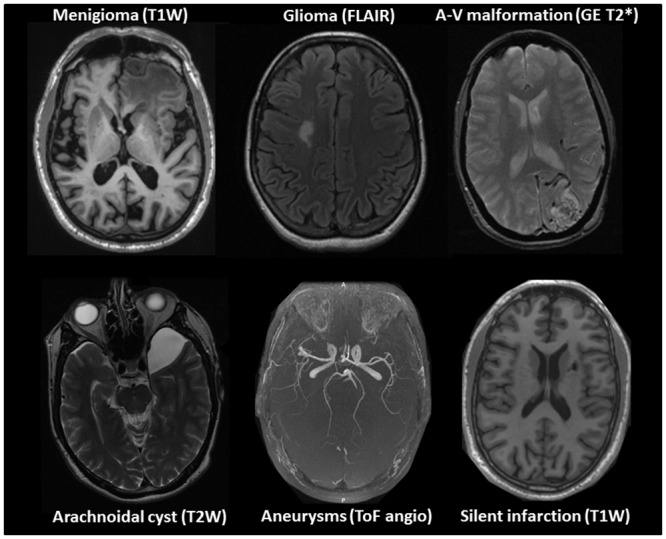
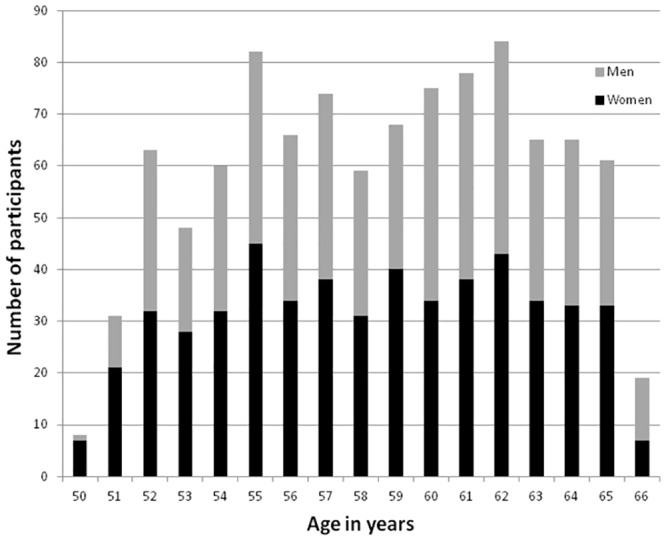
Material and methods: 1006 participants (530 women) underwent MRI of the head at 1.5T consisting of T1 weighted sagittal IR-FSPGR volume, axial T2 weighted, gradient echo T2* weighted and FLAIR sequences plus time of flight cerebral angiography covering the circle of Willis. The nature of a finding and if it was incidental were determined from previous radiological examinations, patient records, phone interview, and/or additional neuroimaging. Handling and outcome of the clinically relevant incidental findings were prospectively recorded. True and false positives were estimated from the repeated neuroimaging.
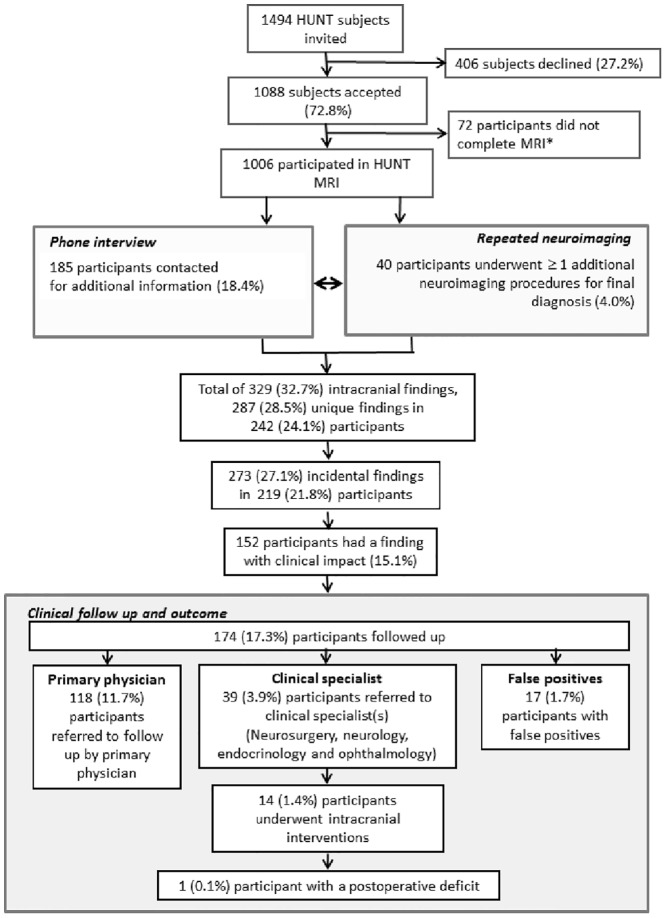
Results: Prevalence of any intracranial finding was 32.7%. Incidental intracranial findings were present in 27.1% and clinically relevant findings in 15.1% of the participants in the HUNT MRI cohort. 185 individuals (18.4%) were contacted by phone about their findings. 40 participants (6.2%) underwent ≥ 1 additional neuroimaging session to establish etiology. Most false positives were linked to an initial diagnosis of suspected glioma, and overall positive predictive value of initial MRI was 0.90 across different diagnoses. 90.8% of the clinically relevant incidental findings were developmental and acquired cerebrovascular pathologies, the remaining 9.2% were intracranial tumors, of which extra-axial tumors predominated. In total, 3.9% of the participants were referred to a clinical specialist, and 11.7% to their primary physician. 1.4% underwent neurosurgery/radiotherapy, and 1 (0.1%) experienced a procedure related postoperative deficit.
Conclusions: In a general population between 50 and 66 years most intracranial findings on MRI were incidental, and >15% of the cohort was referred to clinical-follow up. Hence good routines for handling of findings need to be in place to ensure timely and appropriate handling.
Conflict of interest statement
Figures
References
Publication types
MeSH terms
LinkOut - more resources
Full Text Sources
Other Literature Sources
Medical
