Clinical Trial
. 2016 Apr 1;126(4):1482-94.
doi: 10.1172/JCI85317. Epub 2016 Mar 7.

H7N9 influenza virus neutralizing antibodies that possess few somatic mutations

Clinical Trial

H7N9 influenza virus neutralizing antibodies that possess few somatic mutations

Natalie J Thornburg et al. J Clin Invest. .

Abstract

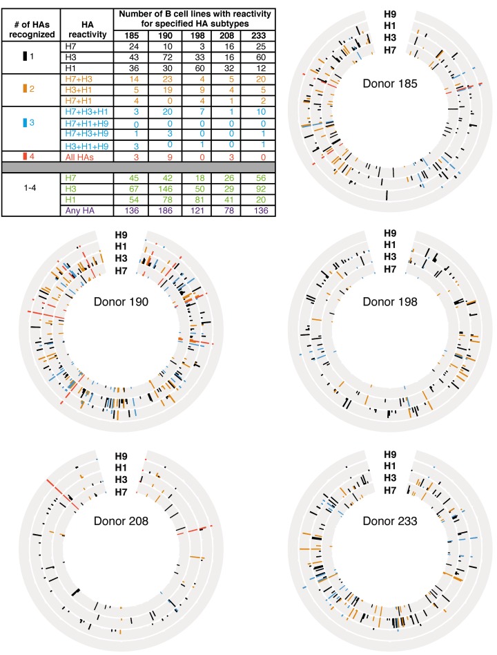
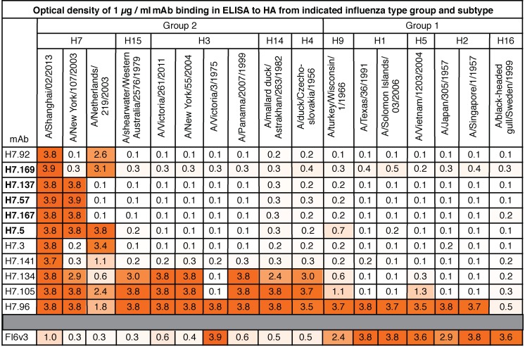
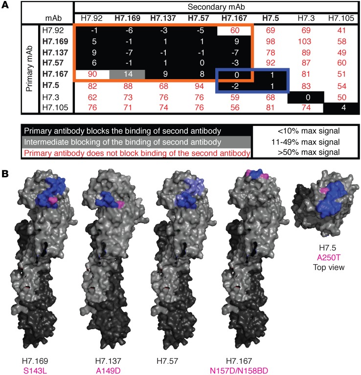
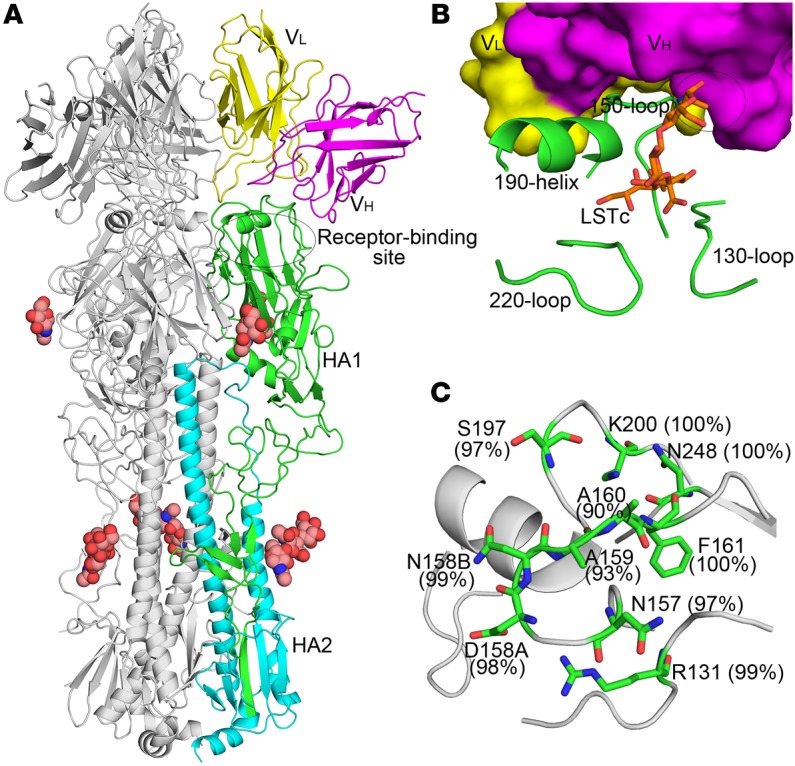
Avian H7N9 influenza viruses are group 2 influenza A viruses that have been identified as the etiologic agent for a current major outbreak that began in China in 2013 and may pose a pandemic threat. Here, we examined the human H7-reactive antibody response in 75 recipients of a monovalent inactivated A/Shanghai/02/2013 H7N9 vaccine. After 2 doses of vaccine, the majority of donors had memory B cells that secreted IgGs specific for H7 HA, with dominant responses against single HA subtypes, although frequencies of H7-reactive B cells ranged widely between donors. We isolated 12 naturally occurring mAbs with low half-maximal effective concentrations for binding, 5 of which possessed neutralizing and HA-inhibiting activities. The 5 neutralizing mAbs exhibited narrow breadth of reactivity with influenza H7 strains. Epitope-mapping studies using neutralization escape mutant analysis, deuterium exchange mass spectrometry, and x-ray crystallography revealed that these neutralizing mAbs bind near the receptor-binding pocket on HA. All 5 neutralizing mAbs possessed low numbers of somatic mutations, suggesting the clones arose from naive B cells. The most potent mAb, H7.167, was tested as a prophylactic treatment in a mouse intranasal virus challenge study, and systemic administration of the mAb markedly reduced viral lung titers.


Figures

Figure 1
Figure 1. Frequency of influenza antigen–specific B cells in H7N9 vaccine recipients.
PBMCs from day 0 (day of vaccination) or day 42 (3 weeks after second vaccination) from 75 donors were transformed with EBV. LCLs were enumerated, and supernatants were assayed for the presence of IgG that bound A/Shanghai/02/2013 H7 HA. The percentage of H7-specific B cells of total transformable B cells was calculated by the number of wells with ODs greater than 2 SDs above background divided by the total number of LCLs times 100. Lines represent the means, and error bars represent SEM. P value was calculated using Wilcoxon test.
Figure 2
Figure 2. Circos plots and summary table representing number of B cell lines with reactivity for specified HAs in 5 donors.
Circos plots from 5 representative donors and a table summary of the results are shown. PBMCs from donors 185, 190, 198, 208, and 233 from day 42 were immortalized with EBV. LCL supernatants were screened by ELISA for the presence of IgGs that bound A/Shanghai/02/2013 or A/New York/55/2004 H7 HAs (H7), A/Hong Kong/1/1968 or A/Victoria/361/2011 H3 HAs (H3), A/California/2009 or A/Texas/36/1991 H1 HAs (H1), or A/turkey/Wisconsin/1/1966 or A/Hong Kong/1073/1999 H9 HA (H9). Each circle has 384 positions, with each representing 1 well of a 384-well plate. Each ring represents results from ELISA using 1 HA protein. The height of the bar represents OD405. Presence of IgG was defined as an OD of greater than 2 SDs above background. Black, gold, aqua or coral bars indicate that a well contained IgG that bound to 1, 2, 3, or 4 HA proteins, respectively. The table represents a summary of data from all 5 Circos plots. The numbers of wells that were reactive against 1 (black), 2 (gold), 3 (aqua), or all 4 (coral) HAs were counted and are indicated in the top portion of the table. The bottom portion of the table represents a tally of all wells with reactivity to any HA and shows the total number of positive wells out of 384 wells tested.
Figure 3
Figure 3. Binding breadth of H7-reactive mAbs.
Binding of each of the 11 H7-reactive mAbs with low EC50 values or the mAb FI6v3 control IgG was tested by ELISA against a panel of recombinant HA molecules at a concentration of 1 μg/ml. Numbers show a mean of OD405 of 4 different wells. Cells are shaded in orange according to OD reading, with higher readings shown as darker. The 5 mAbs previously found to have HAI activity are indicated in bold font.
Figure 4
Figure 4. Epitope mapping of H7-reactive mAbs.
(A) Competition-binding assays were used to bin mAbs into groups that bound the same antigenic regions. Using biolayer interferometry, His-tagged trimeric A/Shanghai/2/2013 H7 HA0 protein was immobilized on Ni-NTA biosensor tips. Two different IgGs then were loaded successively to determine whether the first mAb blocked binding of the second mAb to HA. The first mAb added is listed in the vertical column, and the second mAb added is listed in the horizontal row. The maximal signal of each mAb alone was calculated. The signal of each second mAb was calculated in comparison with that of the mAb alone. mAbs listed in bold font are the mAbs with HAI activity. mAbs in the same competition-binding groups are highlighted in orange and blue boxes. (B) Regions of mAb contact are mapped onto the surface of A/Shanghai/02/2013 H7 HA, as determined by sequence analysis of antibody escape mutant viruses (magenta) or DXMS experiments (blue). The side view is shown for H7.169, H7.137, H7.57, and H7.167. The top view is shown for H7.5. Escape mutant virus HA altered residues are listed below the mAb name in magenta.
Figure 5
Figure 5. The antibody H7.167 binds a conserved epitope adjacent to the RBS of H7 HA.
(A) Crystal structure of H7N9 (A/Shanghai/2/2013, abbreviated as Sh2) HA in complex with H7.167 Fab. One HA/Fab protomer of the trimeric complex is colored with HA1 in green, HA2 in cyan, the Fab heavy chain in magenta, and the Fab light chain in yellow. Only the Fv of the Fab is shown. The other 2 protomers are colored gray. Glycans are shown as spheres (carbon in light pink, oxygen in red, and nitrogen in blue). The constant domain of the Fab cannot be modeled due to disorder that arises from flexibility about the Fab elbow region. (B) Close-up view of the interaction between H7.167 and Sh2/H7 HA. The coloring is similar to that shown in A, but H7.167 is shown in a surface representation. The human receptor analog LSTc (orange sticks) was docked into the RBS of the complex by its superimposition with human receptor analog from human H7 HA–LSTc complex (PDB ID: 4BSE). The RBS elements (130-loop, 150-loop, 190-helix, and 220-loop) are also labeled. The variable domain of the heavy chain (VH) shows an obvious clash (labeled as an ellipse) with 1 glycan of LSTc. (C) Sequence conservation of the H7.167 footprint across H7 viruses. HA1 is shown in gray, and the epitope residues are shown as green sticks. The weighted percentage of identity of each residue with the H7 consensus sequence is indicated.
Figure 6
Figure 6. Interaction of H7.167 with Sh2/H7 illustrating the neutralizing epitope.
(A) Surface presentation of H7.167 footprint (green) in Sh2/H7 HA (gray) highlighting antibody residues contacting HA. The relative location of RBS is labeled. (B) The electrostatic potential surface of HA is illustrated (red, negative, –5.4 kT; blue, positive, +5.4 kT; white, neutral) in the same orientation as in A, with the H7.167 contacting residues in the 5 CDRs that interact with HA represented as sticks and color coded by CDR. H7.167 binds HA with both chains, using 5 of the 6 CDRs (LCDR2 makes no contacts) of the heavy and light chains in recognition (magenta and yellow, respectively).
Figure 7
Figure 7. Viral lung titers and weight from mice treated with H7.167.
Mice were injected with an irrelevant mAb, BDBV280, or H7.167 at 1.65 mg/kg or 5 mg/kg i.p. 1 day prior to viral inoculation. Mice were anesthetized and inoculated with 10,000 PFU H7-PR8 reassortant virus i.n. and were euthanized on day 3 (n = 3) or 5 (n = 3) to measure the presence of virus in lung tissue (A) or weighed every day for 2 weeks (n = 5) (B). P value was calculated using an unpaired t test with equal SD.
Figure 8
Figure 8. Mutation frequency of influenza mAbs.
(A) Antibody variable gene sequences were determined and analyzed using the IMGT analysis tool. The percentage of identity of antibody heavy-chain variable genes as compared with inferred germline genes is shown for H7-specific mAbs with or without HAI activity. Nucleotide mutation frequencies are shown and represent both coding and noncoding mutations. Lines represent the means, and error bars represent SEM. P values were calculated using the Mann-Whitney U test. (B) The total number of amino acid mutations in heavy and light chains as compared with inferred germline genes, as determined by IMGT analysis. Genes encoding H7-specific or H7 mAbs with heterosubtypic binding activity from this manuscript were compared with sequences of previously published H5-specific, H1-specific, or broadly neutralizing influenza mAbs. The lines represent the means, and error bars represent SEM. P values were calculated using the Mann-Whitney U test.

References

    1. World Health Organization WHO RISK ASSESSMENT of Human infections with avian influenza A(H7N9) virus. [February 23, 2015]; [February 1, 2016];WHO website. http://www.who.int/influenza/human_animal_interface/influenza_h7n9/RiskA...
    1. Gao R, et al. Human infection with a novel avian-origin influenza A (H7N9) virus. N Engl J Med. 2013;368(20):1888–1897. doi: 10.1056/NEJMoa1304459. - DOI - PubMed
    1. Liu D, et al. Origin and diversity of novel avian influenza A H7N9 viruses causing human infection: phylogenetic, structural, and coalescent analyses. Lancet. 2013;381(9881):1926–1932. doi: 10.1016/S0140-6736(13)60938-1. - DOI - PubMed
    1. Wang Y, et al. Towards a better understanding of the novel avian-origin H7N9 influenza A virus in China. Sci Rep. 2013;3: - PMC - PubMed
    1. Kageyama T, et al. Genetic analysis of novel avian A(H7N9) influenza viruses isolated from patients in China, February to April 2013. Euro Surveill. 2013;18(15): - PMC - PubMed

Publication types

MeSH terms