. 2016 Apr;34(4):424-9.
doi: 10.1038/nbt.3513. Epub 2016 Mar 7.

Targeted gene addition in human CD34(+) hematopoietic cells for correction of X-linked chronic granulomatous disease

Affiliations

Targeted gene addition in human CD34(+) hematopoietic cells for correction of X-linked chronic granulomatous disease

Suk See De Ravin et al. Nat Biotechnol. 2016 Apr.

Abstract

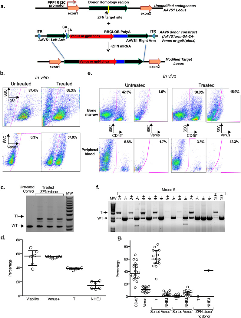
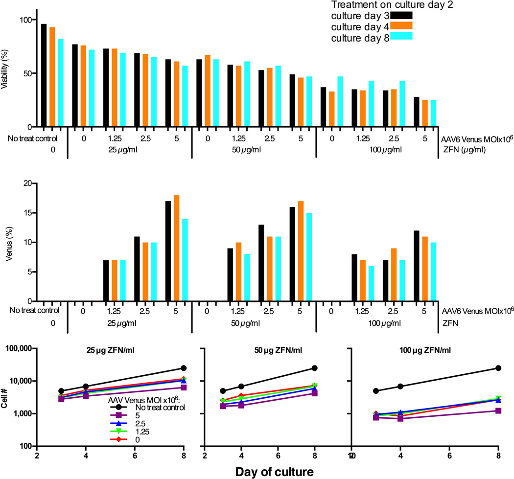
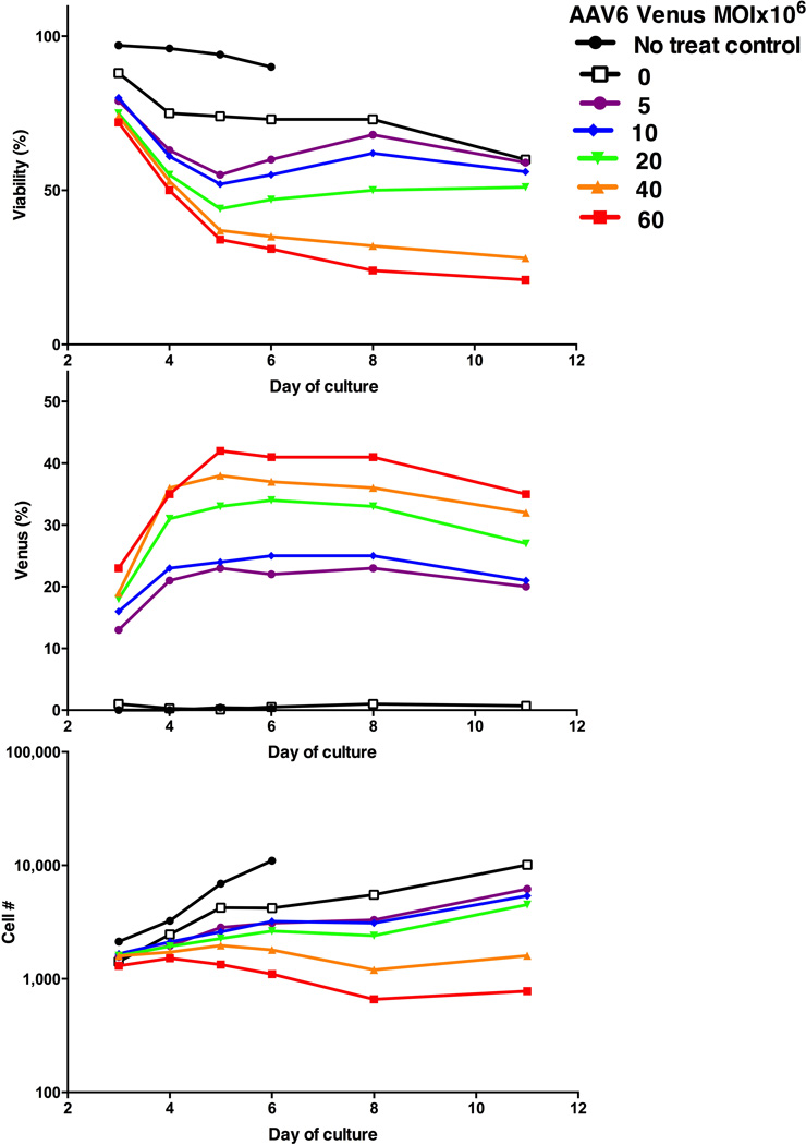
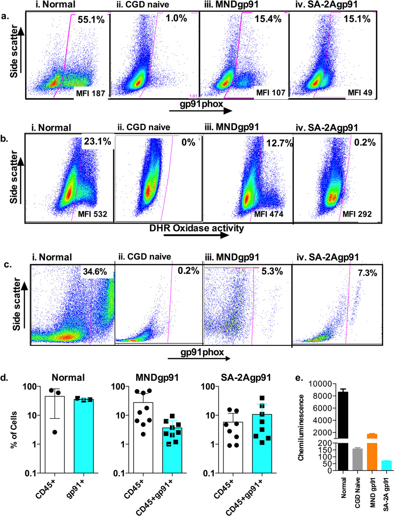
Gene therapy with genetically modified human CD34(+) hematopoietic stem and progenitor cells (HSPCs) may be safer using targeted integration (TI) of transgenes into a genomic 'safe harbor' site rather than random viral integration. We demonstrate that temporally optimized delivery of zinc finger nuclease mRNA via electroporation and adeno-associated virus (AAV) 6 delivery of donor constructs in human HSPCs approaches clinically relevant levels of TI into the AAVS1 safe harbor locus. Up to 58% Venus(+) HSPCs with 6-16% human cell marking were observed following engraftment into mice. In HSPCs from patients with X-linked chronic granulomatous disease (X-CGD), caused by mutations in the gp91phox subunit of the NADPH oxidase, TI of a gp91phox transgene into AAVS1 resulted in ∼15% gp91phox expression and increased NADPH oxidase activity in ex vivo-derived neutrophils. In mice transplanted with corrected HSPCs, 4-11% of human cells in the bone marrow expressed gp91phox. This method for TI into AAVS1 may be broadly applicable to correction of other monogenic diseases.


Figures

Figure 1
Figure 1. AAVS1 specific ZFN and AAV6 donor mediated targeted insertion of Venus fluorescent marker into human CD34+ HSCs
a) A schematic of the AAVS1 locus (top) indicating the ZFN target site within PPP1R12C and the AAV6 donor homology regions (green). The AAV6 donor construct (middle) contains homology sequences flanking the Venus marker or gp91phox therapeutic cDNA (red) preceded by a splice-acceptor (SA) and a 2A cleavage element, and followed by a rabbit globin polyA signal. The TI modified locus is depicted below. Not shown is the gp91phox donor construct in which SA-2A is replaced by the MND promoter. b) Flow cytometry analysis of Venus expression in 10 day cultured human HSCs with untreated or gene targeted cells in left and right panels, respectively. Treated cells were targeted by electroporation of ZFN mRNA (25µg/ml each) and transduction with AAV6-SA-2A-Venus (1×10⁶ MOI) on day 2 of culture. Top panels show gating of viable cells analyzed for Venus expression in the bottom panels. c) Electrophoresis gel molecular analysis of TI at the AAVS1 locus in HSCs treated as in Fig 1b. PCR with primers outside the homology region (HDR-F4 and HDR-R5 in Suppl. Fig. 5a) result in a 3.1kb product of targeted Venus insertion (TI) only seen in treated HSCs (n=3), or a 2kb product representing endogenous locus (wild type; WT). d) Bar graph summary of in vitro efficiency of Venus TI (n=6) showing flow cytometry analysis as analyzed in Fig 1b for viability and Venus expression or showing molecular analysis using MiSeq of TI versus NHEJ at the AAVS1 site in treated HSCs as described in detail in the Methods. e) Flow cytometry analysis of Venus expression in bone marrow (top panels) or peripheral blood (bottom panels) from NSG mice 17 weeks after transplant of human HSCs treated as in Fig 1b, but with average Venus expression of 30% before transplant. Shown are results from mice transplanted with untreated human HSCs (left panel pairs) or Venus TI treated HSCs (right panel pairs). The left panel of each pair shows gating of human CD45+ cells analyzed for Venus expression in the right panel of each pair. f) Electrophoresis gel molecular analysis of human HSCs harvested from bone marrow of 17-week post-transplant NSG mice treated as described in Fig 1e and using primers for analysis of TI at the AAVS1 locus as described in Fig 1c. Marrow from each mouse was sorted into a human CD45+ Venus+ fraction (“+” after each mouse # at top of gel lane) and human CD45+ Venus fractions “−“) for TI analysis. 16 mice were transplanted with human HSCs treated with Venus TI of which analysis of 8 is shown in this gel (Mice 1–7 and 10). Two mice were transplanted with control human HSCs, where mouse #8 received HSCs electroporated with AAVS1 ZFNs without AAV6 Venus donor, while mouse #9 received HSCs treated with AAV6 Venus donor alone (#9). No Venus expression was detected by flow cytometry in marrow cells from the controls, so only TI analysis of the human CD45+ Venus sorted fractions were analyzed. g) Bar graph summary of flow cytometry analysis and molecular analysis of Venus transgene in bone marrow from the NSG mice 17 weeks after transplant of human HSCs treated and analyzed as in Fig 1e and 1F. The first 2 bars from the left show, respectively, the level of human cell engraftment in mice transplanted with Venus TI treated human HSCs and percent of Venus expression in the human cell graft (n=16). Following cell sorting of marrow as described in Fig 1f the Venus+ (sorted V+) and Venus (sorted V) fractions (middle 4 bars) were analyzed by MiSeq for presence of Venus transgene (T) and for NHEJ (n=14). The two bars at the right side of the graph show analysis of the control mouse transplanted with human HSCs treated with ZFN without AAV6 Venus donor, leading to high NHEJ without any detectable transgene. The quantification of Venus transgene and NHEJ was assessed using only the MiSeq primer set (Mi-F,Mi-R, plus 2A-R), without first amplifying with an Out/Out primer set (see Suppl. Fig 5a and as described in detail in the Methods). Thus, this primer set detects donor both within AAVS1 and off-target anywhere in the genome (T).
Figure 2
Figure 2. Optimization of ratios of AAVS1 ZFN mRNA to AAV6 Venus donor
On day two of culture healthy donor CD34+ HSCs (2×10⁵ cells/10 µl per sample) were electroporated (MaxCyte GT instrument) with a series of indicated AAVS1 ZFN mRNA pair concentrations immediately followed by exposure to indicated AAV6 Venus donor MOIs. This figure shows cell viability (top), percent of live cells expressing Venus marker (middle) and relative viable cell numbers at each day of culture after treatment in each sample following treatment to reflect cell proliferative capability after treatment (bottom). Values were determined for each analysis at culture day 3, 4 and 8 (corresponding to 1, 2 and 6 days post treatment).
Figure 3
Figure 3. Optimization of AAV6 Venus donor MOI
Healthy donor CD34+ HSCs (2×10⁵ cells/10 µl per sample) were electroporated with AAVS1 ZFN mRNAs at 25g/mL, followed by transduction with the indicated dilutions of AAV6 Venus. This figure shows cell viability (top), percent of live cells expressing Venus marker (middle) and relative viable cell numbers (bottom) at indicated days of culture after treatment in each sample.
Figure 4
Figure 4. AAVS1 specific ZFN and AAV6 donor mediated targeted insertion of gp91phox corrective gene into human CD34+ HSCs from patients with X-linked chronic granulomatous disease (X-CGD)
a) Flow cytometry analysis of gp91phox expression in 7 day cultured untreated healthy human donor HSCs (normal), untreated XCGD patient HSCs (CGD naïve), ZFN plus AAV6-MND-gp91 TI treated XCGD patient HSCs (MNDgp91), and ZFN plus AAV6-SA-2A-gp91 TI treated XCGD patient HSCs (SA-2Agp91). Indicated at the top of the gated area of each panel is the percent of gp91phox positive cells, where the mean fluorescence intensity (MFI) indicates the average per cell expression of gp91phox positive cell. b) Flow cytometry dihydrorhodamine (DHR) analysis of NADPH oxidase activity in 14 day cultured untreated healthy human donor HSCs (normal), untreated XCGD patient HSCs (CGD naïve), ZFN plus AAV6-MND-gp91 TI treated XCGD patient HSCs (MNDgp91), and ZFN plus AAV6-SA-2A-gp91 TI treated XCGD patient HSCs (SA-2Agp91). Indicated at the top of the gated area of each panel is the percent of oxidase positive cells, where the mean fluorescence intensity (MFI) indicates the average per cell oxidase activity per positive cell. c) Flow cytometry analysis of gp91phox expression of the human CD45+ cells in bone marrow from NSG mice 8 weeks after transplant of untreated healthy human donor HSCs (normal), untreated XCGD patient HSCs (CGD naïve), ZFN plus AAV6-MND-gp91 TI treated XCGD patient HSCs (MNDgp91), and ZFN plus AAV6-SA-2A-gp91 TI treated XCGD patient HSCs (SA-2Agp91). The gp91phox expression in culture cells before transplant is as shown in Fig 2a. Indicated at the top of the gated area of each panel is the percent of gp91phox positive cells. d) Bar graph summary of flow cytometry analysis of the level of human cell engraftment (CD45+) and percent of the human CD45+ cells expressing gp91phox (CD45+gp91+) in bone marrow of mice transplanted with untreated healthy human donor HSCs (normal) (n=3), ZFN plus AAV6-MND-gp91 TI treated XCGD patient HSCs (MNDgp91) (n=9), and ZFN plus AAV6-SA-2A-gp91 TI treated XCGD patient HSCs (SA-2Agp91) (n=9). The average % is shown at the top of each bar. e) Functional correction in X-CGD patient CD34+ progenitors. Bar graph summary of phorbol myristate acetate stimulated chemiluminescence detection of reactive oxidative species production from similarly in vitro cultured CD34+ progenitors differentiated into myeloid cells over × days in culture from a normal donor (N), untreated XCGD patient HSCs (CGD naïve), ZFN plus AAV6-MND-gp91 TI treated XCGD patient HSCs (MNDgp91), and ZFN plus AAV6-SA-2A-gp91 TI treated XCGD patient HSCs (SA-2Agp91). The mean chemiluminescence intensity units are shown at the top of each bar.

References

    1. Biffi A, et al. Lentiviral hematopoietic stem cell gene therapy benefits metachromatic leukodystrophy. Science. 2013;341:1233158. - PubMed
    1. Hacein-Bey-Abina S, et al. Efficacy of gene therapy for X-linked severe combined immunodeficiency. N Engl J Med. 2010;363:355–364. - PMC - PubMed
    1. Cavazzana-Calvo M, et al. Gene therapy of human severe combined immunodeficiency (SCID)-X1 disease. Science. 2000;288:669–672. - PubMed
    1. Hacein-Bey-Abina S, et al. A serious adverse event after successful gene therapy for X-linked severe combined immunodeficiency. The New England journal of medicine. 2003;348:255–256. - PubMed
    1. Stein S, et al. Genomic instability and myelodysplasia with monosomy 7 consequent to EVI1 activation after gene therapy for chronic granulomatous disease. Nature medicine. 2010;16:198–204. - PubMed

Publication types