. 2016 Jul;18(7):939-49.
doi: 10.1093/neuonc/now005. Epub 2016 Mar 6.

Decrease of VEGF-A in myeloid cells attenuates glioma progression and prolongs survival in an experimental glioma model

Affiliations

Decrease of VEGF-A in myeloid cells attenuates glioma progression and prolongs survival in an experimental glioma model

Nadja Osterberg et al. Neuro Oncol. 2016 Jul.

Abstract

Background: Glioblastomas are highly vascularized tumors with a prominent infiltration of macrophages/microglia whose role in promoting glioma growth, invasion, and angiogenesis has not been fully elucidated.

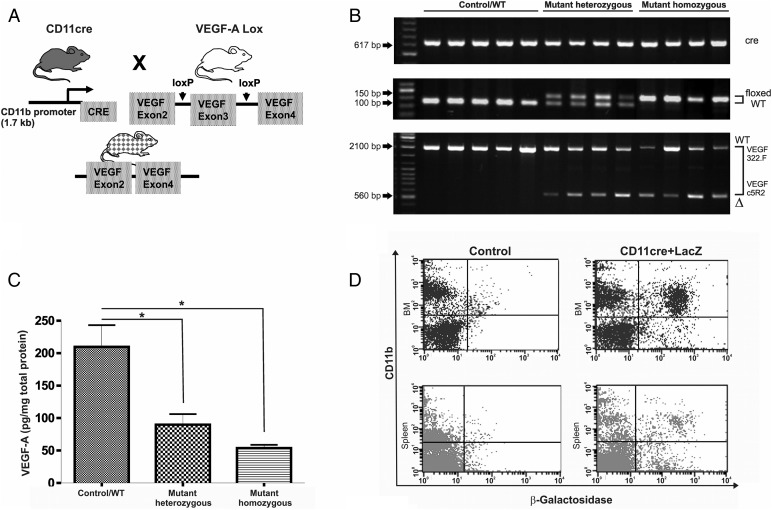
Methods: The contribution of myeloid-derived vascular endothelial growth factor (VEGF) to glioma growth was analyzed in vivo in a syngeneic intracranial GL261 glioma model using a Cre/loxP system to knock out the expression of VEGF-A in CD11b + myeloid cells. Changes in angiogenesis-related gene expression profile were analyzed in mutant bone marrow-derived (BMD) macrophages in vitro. Furthermore, we studied the influence of macrophages on GL261 growth, invasiveness, and protein expression profile of angiogenic molecules as well as the paracrine effect of mutant macrophages on angiogenesis in vitro.

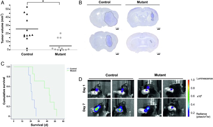
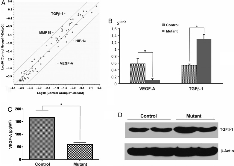
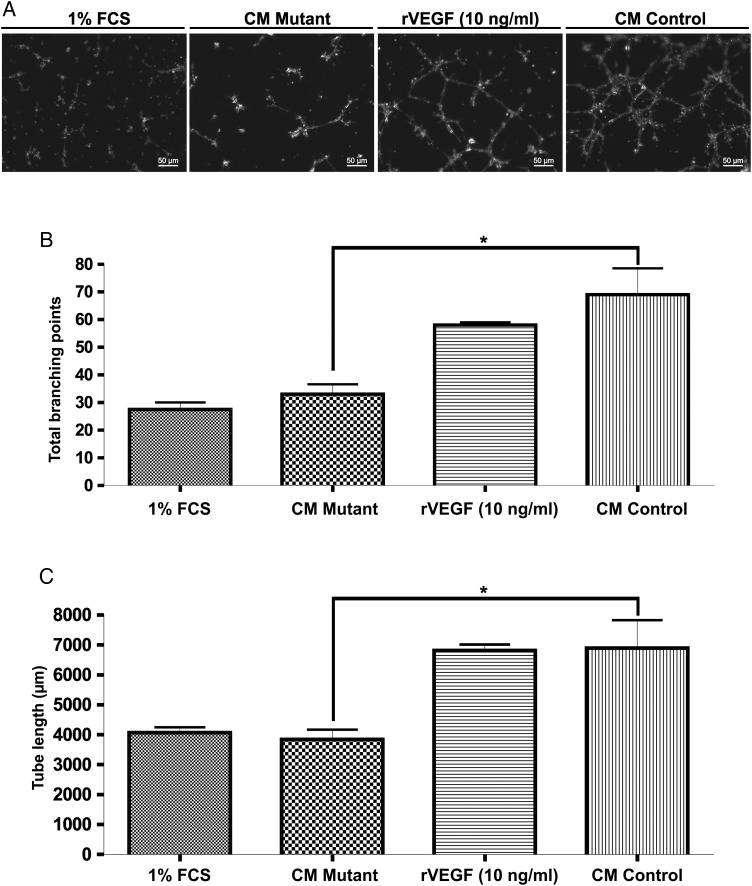
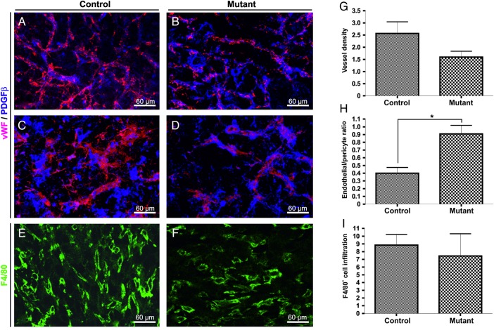
Results: Myeloid cell-restricted VEGF-A deficiency leads to a growth delay of intracranial tumors and prolonged survival. The tumor vasculature in mutant mice was more regular, with increased pericyte coverage. Expression analysis revealed significant downregulation of VEGF-A and slight upregulation of TGFβ-1 in BMD macrophages from mutant mice. Endothelial tube formation was significantly decreased by conditioned media from mutant macrophages. The expression of angiogenesis-related proteins in GL261 glioma cells in co-culture experiments either with wild-type or mutant macrophages remained unchanged, indicating that effects observed in vivo are due to myeloid-derived VEGF-A deficiency.

Conclusions: Our results highlight the importance of VEGF derived from tumor-infiltrating myeloid cells for initiating vascularization in gliomas. The combination of antiangiogenic agents with myeloid cell-targeting strategies might provide a new therapeutic approach for glioblastoma patients.

Keywords: VEGF; angiogenesis; glioma; macrophages; microglia.


Figures

Fig. 1.
Fig. 1.
Characterization of CD11bcre + VEGFlox/lox mice. (A) Schematic diagram of the transgenic mice used to generate mutant CD11bcre + VEGFlox/lox. (B) Mice genotyping: a band of 617 bp indicates the presence of cre transgene (top panel). A 100 bp band indicates wild-type (WT) VEGF-A allele; a 150 bp band corresponds to homozygous floxed VEGF-A (mutant homo VEGFlox/lox) allele. Heterozygous mice showed 100 bp and 150 bp bands (mutant hetero VEGF+/lox) (middle panel). PCR amplification of genomic DNA from bone marrow-derived (BMD) macrophage preparations with primers that bind to either side of VEGF-A exon 3 demonstrate the partial excision of this stretch of DNA. (A band of 2.1 kb represents wild-type VEGF-A+/+, and a band of 560 bp represents mutant mice (bottom panel). (C) Detection of VEGF-A in supernatants of cultured BMD-macrophages (control n = 9; mutant hetero n = 4; mutant homo n = 5). Bars represent means and ± SEM, (*P < .05) (D) Quantification of expression of LacZ reporter driven by the CD11bcre promoter in bone marrow and spleen cells derived from CD11bcre + LacZ mice. Fluorescence-activated cell sorting (FACS) analysis disclosed that 40% of the CD11b+ cells in bone marrow expressed the reporter gene. Low levels of recombination were detected in spleen macrophages.
Fig. 2.
Fig. 2.
Myeloid-derived VEGF-A plays an important role in glioma growth. (A) Analysis of tumor volumes in control (CD11bcre + VEGFA+/+) and in mutant (CD11bcre + VEGFlox/lox) mice. Results for individual animals are present. The bold lines represent the mean in each group (*P < .05). (B) Representative images of brain section of intracranial tumor-bearing mice. Mice were euthanized at day 18 post tumor implantation. Sections (50 µm) were stained with hematoxylin . Scale bar = 1 mm. (C) Kaplan-Meier survival analysis of mice implanted with GL261 stably expressing luciferase showing the prolonged survival of mutant mice. (D) Localization of bioluminescence (BLI) signal at day 1 and day 7 post implantation of GL261 expressing luciferase. Representative BLI scans show the anatomical injection site.
Fig. 3.
Fig. 3.
Gene expression profile of in vitro differentiated macrophages. (A) Plot of a panel of 84 angiogenesis-related genes with a fold-change boundary >5. The differential expression of in vitro differentiated macrophages derived from control versus mutant mice (n = 4/group) was evaluated using the RT2 Profiler Array System. VEGF-A and HIF-1α were downregulated, whereas MMP19 and TGFb-1 were upregulated. (B) Validation of PCR array screening using real-time PCR confirmed the differential regulation of VEGF-A and TGFb-1 at mRNA levels. (C) VEGF-A ELISA of supernatants from control and mutant macrophages confirms that VEGF-A protein in mutant macrophages was significantly decreased but not abolished. Bars represent means ± SEM, (*P < .05). (D) Representative Western blot analysis of TGFβ-1 protein expression in bone marrow-derived macrophages. Densitometric quantification of 3 independent experiments revealed a 1.5 increase of TGFβ-1 in mutant macrophages.
Fig. 4.
Fig. 4.
Decreased VEGF-A expression by macrophages inhibits tube formation in vitro. (A) Representative images of Matrigel tube formation assay after a 12 hour incubation. Rat brain endothelial cells were incubated with conditioned media from control and mutant macrophages. Media containing 1% FCS and recombinant mouse VEGF-A were used as control. The graphs display total branching points (B) and the total tube length (C) (2 samples/group). Bars represent means ± SEM, (*P < .05).
Fig. 5.
Fig. 5.
Normalization of the tumor vasculature gliomas grown in mutant mice. Representative confocal microscopy images of double immunofluorescence for von Willebrand's factor( vWF; detection of endothelial cells, red) and PDGFRβ (detection of pericytes, blue) showing the vascular network in intracranial gliomas from control (A and C) and mutant animals (B and D). Quantitative analysis of vascular density (G) and pericyte cover index (H) in the different groups (n = 3–5 animals/group). Representative confocal images of macrophage infiltration determined by staining with F4/80 in control (E) and mutant (F) tumors. Quantification analysis shows no substantial difference in the accumulation of inflammatory cells in mutant tumors compared with control tumors (I). Scale bar = 30 µm. Bars represents means ± SEM, (*P < .05).
Fig. 6.
Fig. 6.
Infiltrating tumor macrophages are a source of VEGF in human glioblastomas. Human glioblastoma serial sections were stained immunohistochemically with anti-VEGF (D and E) anti-CD68 (A and B), anti-CD14 (C), and anti-CD34 (F). High magnification of boxed areas is shown in B and C. Both tumor cells and macrophages produce VEGF in glioblastomas. VEGF protein is found on endothelial cells of CD34 + tumor vessels and particularly in perivascular CD68 + macrophages. Scale bar 50 µm.

References

    1. Machein M, de Miguel LS. Angiogenesis in gliomas. Recent Results Cancer Res. 2009;171:193–215. - PubMed
    1. Machein MR, Plate KH. VEGF in brain tumors. J Neurooncol. 2000;50(1–2):109–120. - PubMed
    1. Allavena P, Mantovani A. Immunology in the clinic review series; focus on cancer: tumour-associated macrophages: undisputed stars of the inflammatory tumour microenvironment. Clin Exp Immunol 2012;167(2):195–205. - PMC - PubMed
    1. Mantovani A, Germano G, Marchesi F, Locatelli M, Biswas SK. Cancer-promoting tumor-associated macrophages: new vistas and open questions. Eur J Immunol. 2011;41(9):2522–2525. - PubMed
    1. Zhai H, Heppner FL, Tsirka SE. Microglia/macrophages promote glioma progression. Glia 2011;59(3):472–485. - PMC - PubMed

Publication types

Substances