

Sleep Duration and Subsequent Cortical Thinning in Cognitively Normal Older Adults
- PMID: 26951390
- PMCID: PMC4835311
- DOI: 10.5665/sleep.5768
Sleep Duration and Subsequent Cortical Thinning in Cognitively Normal Older Adults
Abstract
Study objectives: To determine the association between self-reported sleep duration and cortical thinning among older adults.
Methods: We studied 122 cognitively normal participants in the Baltimore Longitudinal Study of Aging with a mean age = 66.6 y (range, 51-84) at baseline sleep assessment and 69.5 y (range, 56-86) at initial magnetic resonance imaging (MRI) scan. Participants reported average sleep duration and completed a mean of 7.6 1.5-T MRI scans (range, 3-11), with mean follow-up from initial scan of 8.0 y (range, 2.0-11.8).
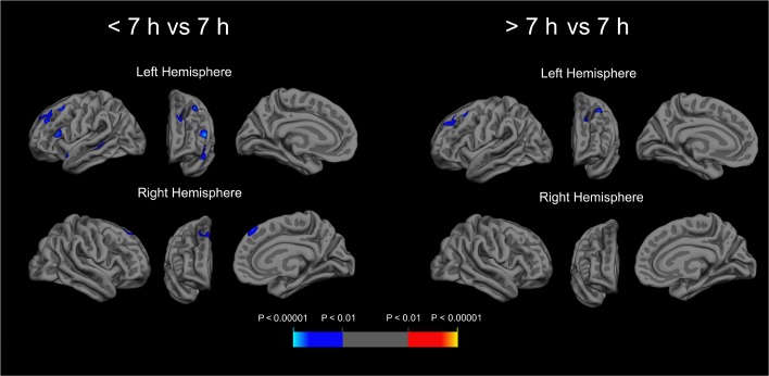
Results: In analyses adjusted for age, sex, education, race, and interval between sleep assessment and initial MRI scan, participants reporting > 7 h sleep at baseline had thinner cortex in the inferior occipital gyrus and sulcus of the left hemisphere at initial MRI scan than those reporting 7 h (cluster P < 0.05). In adjusted longitudinal analyses, compared to those reporting 7 h of sleep, participants reporting < 7 h exhibited higher rates of subsequent thinning in the superior temporal sulcus and gyrus, inferior and middle frontal gyrus, and superior frontal sulcus of the left hemisphere, and in the superior frontal gyrus of the right hemisphere; those reporting > 7 h of sleep had higher rates of thinning in the superior frontal and middle frontal gyrus of the left hemisphere (cluster P < 0.05 for all). In sensitivity analyses, adjustment for apolipoprotein E (APOE) e4 genotype reduced or eliminated some effects but revealed others. When reports of < 7 h of sleep were compared to reports of 7 or 8 h combined, there were no significant associations with cortical thinning.
Conclusions: Among cognitively normal older adults, sleep durations of < 7 h and > 7 h may increase the rate of subsequent frontotemporal gray matter atrophy. Additional studies, including those that use objective sleep measures and investigate mechanisms linking sleep duration to gray matter loss, are needed.
Keywords: aging; atrophy; cortical thinning; gray matter; neuroimaging; sleep duration.
© 2016 Associated Professional Sleep Societies, LLC.
Figures
References
-
- Ohayon MM, Vecchierini MF. Normative sleep data, cognitive function and daily living activities in older adults in the community. Sleep. 2005;28:981–9. - PubMed
-
- Keage HA, Banks S, Yang KL, Morgan K, Brayne C, Matthews FE. What sleep characteristics predict cognitive decline in the elderly? Sleep Med. 2012;13:886–92. - PubMed
-
- Faubel R, Lopez-Garcia E, Guallar-Castillon P, Graciani A, Banegas JR, Rodriguez-Artalejo F. Usual sleep duration and cognitive function in older adults in Spain. J Sleep Res. 2009;18:427–35. - PubMed
Publication types
MeSH terms
Substances
Grants and funding
LinkOut - more resources
Full Text Sources
Other Literature Sources
Medical
Miscellaneous
