. 2016 Mar 8:5:e12509.
doi: 10.7554/eLife.12509.

Trifunctional cross-linker for mapping protein-protein interaction networks and comparing protein conformational states

Affiliations

Trifunctional cross-linker for mapping protein-protein interaction networks and comparing protein conformational states

Dan Tan et al. Elife. .

Abstract

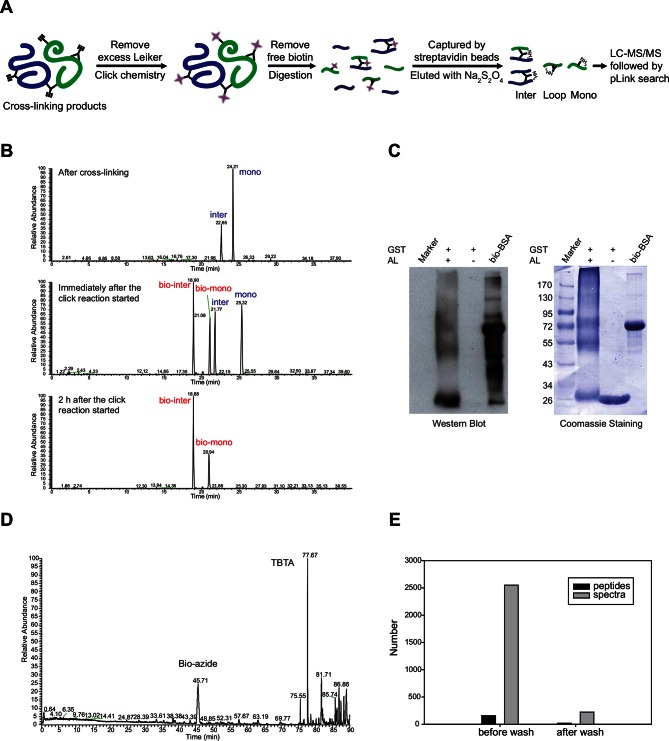
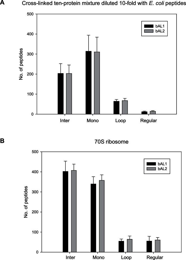
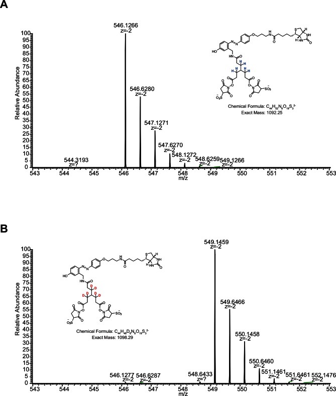
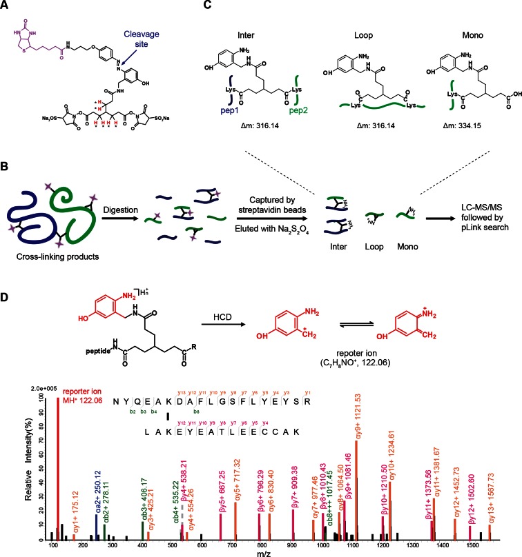
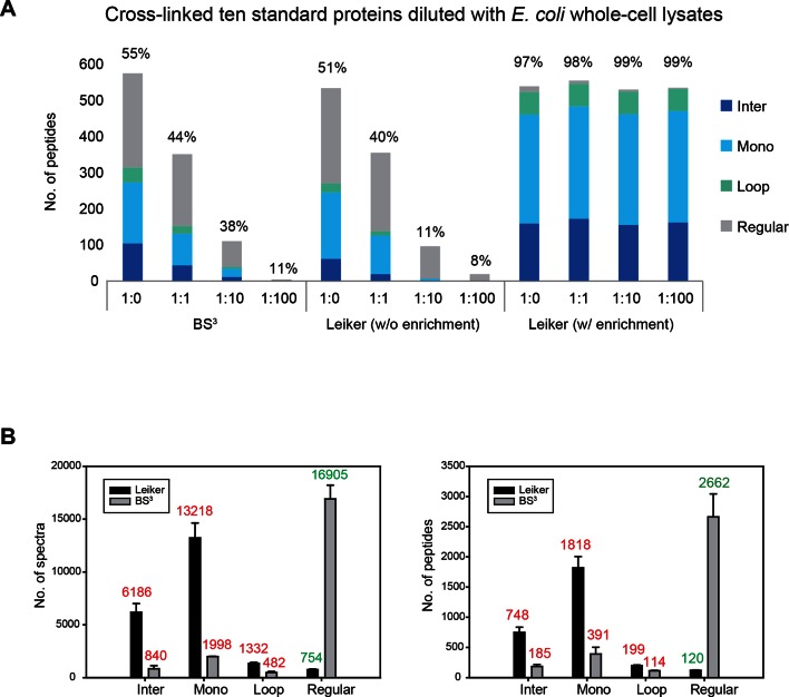
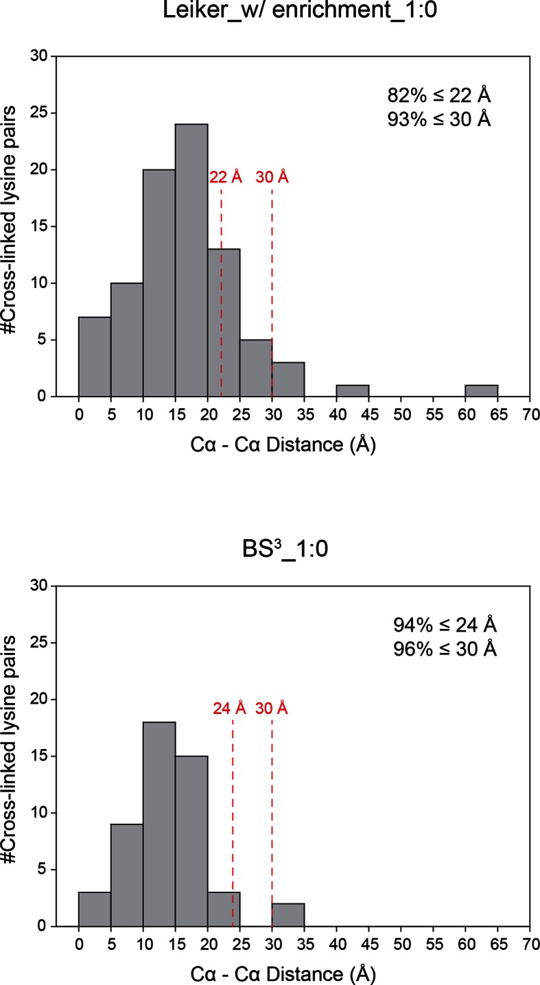
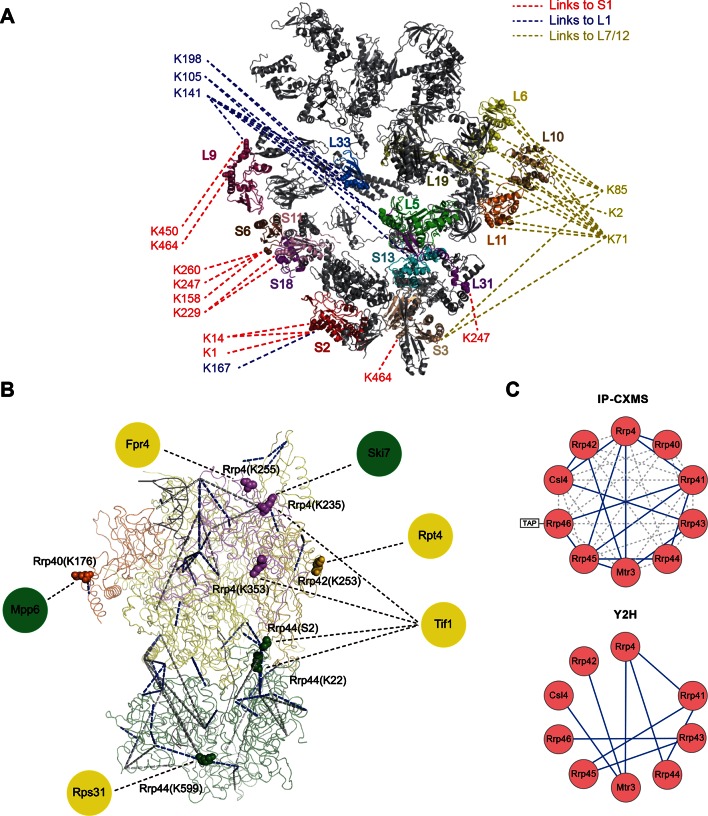
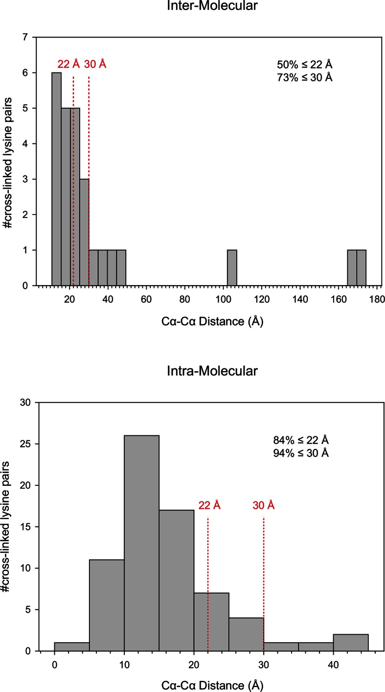
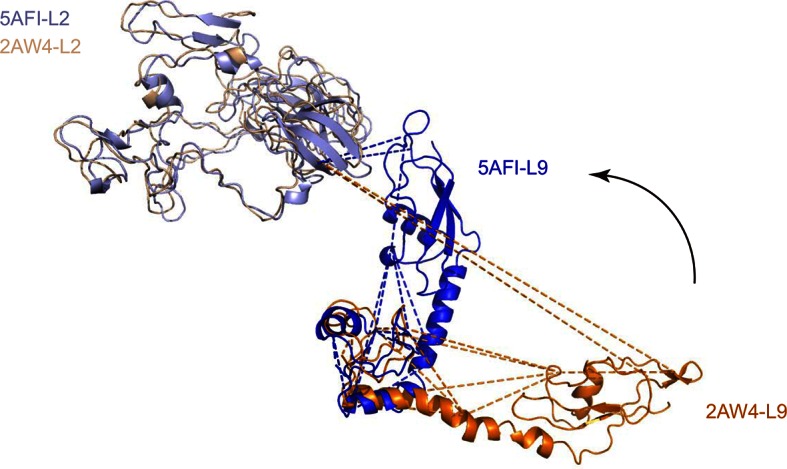
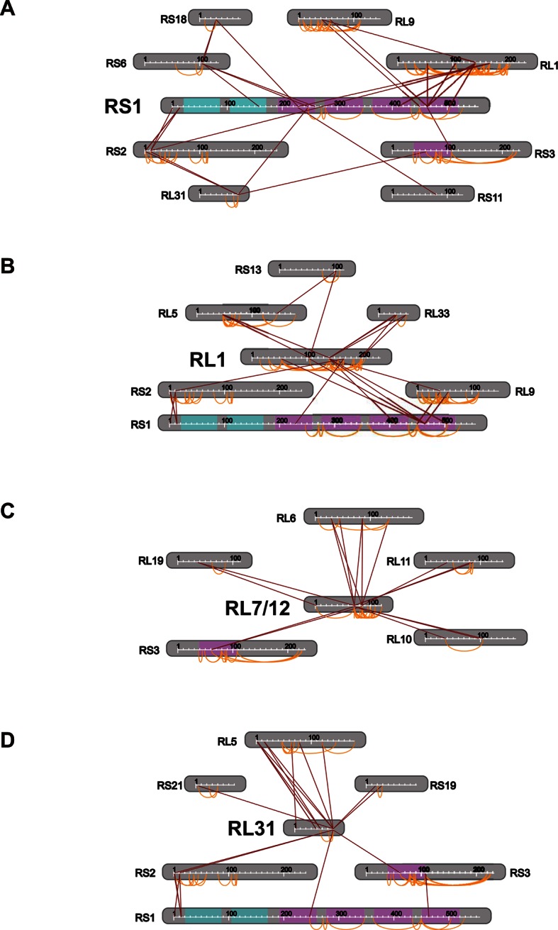
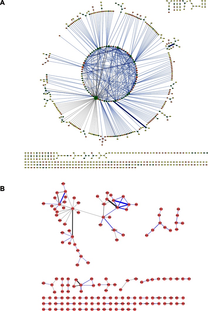
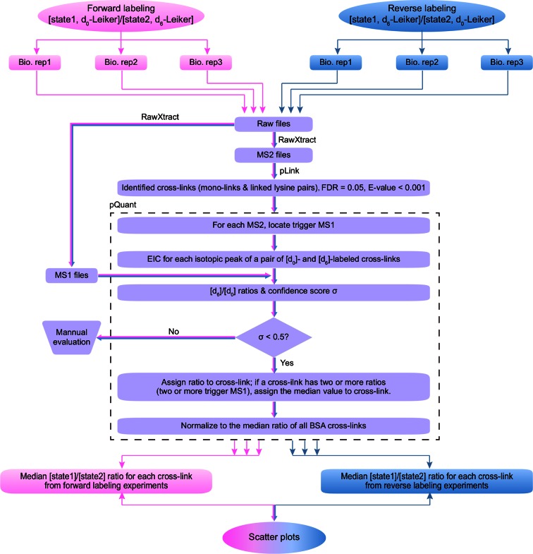
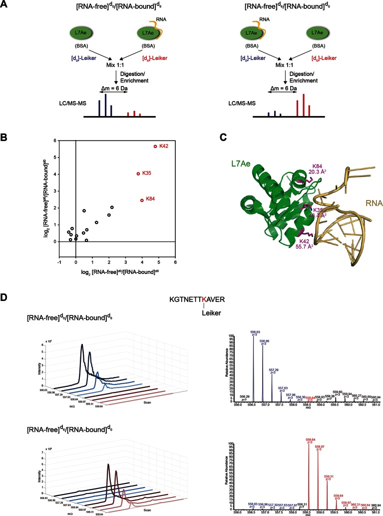
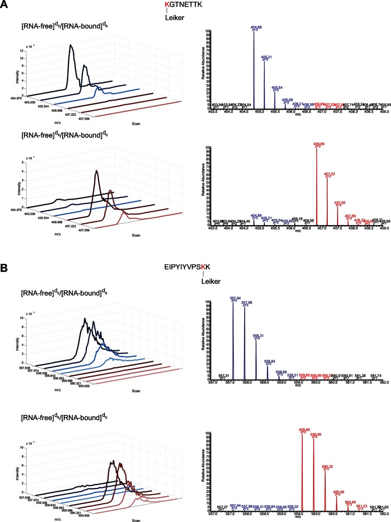
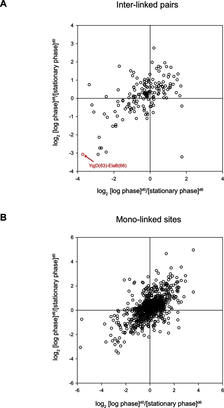
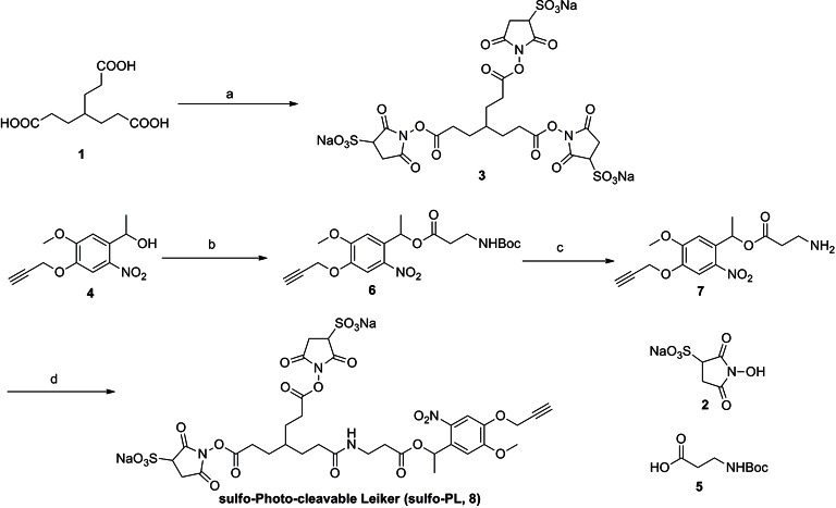
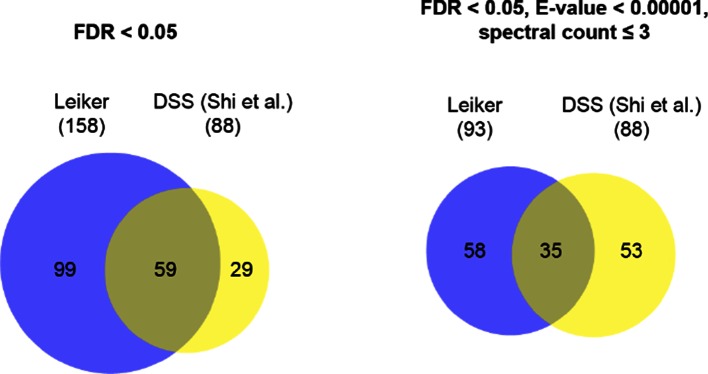
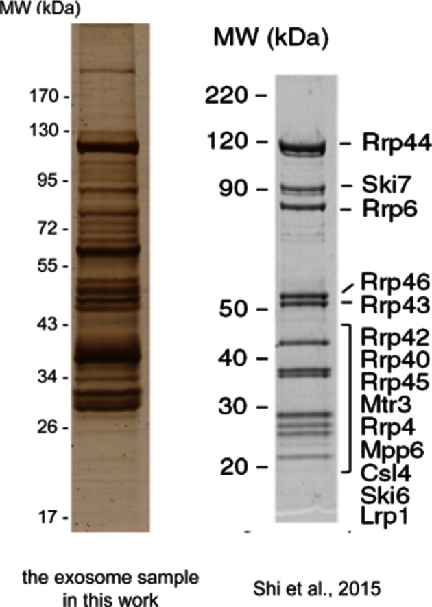
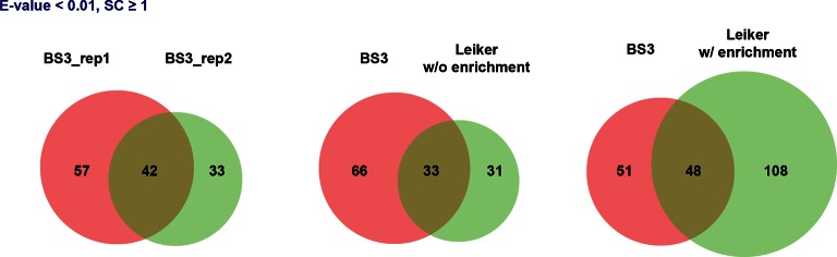
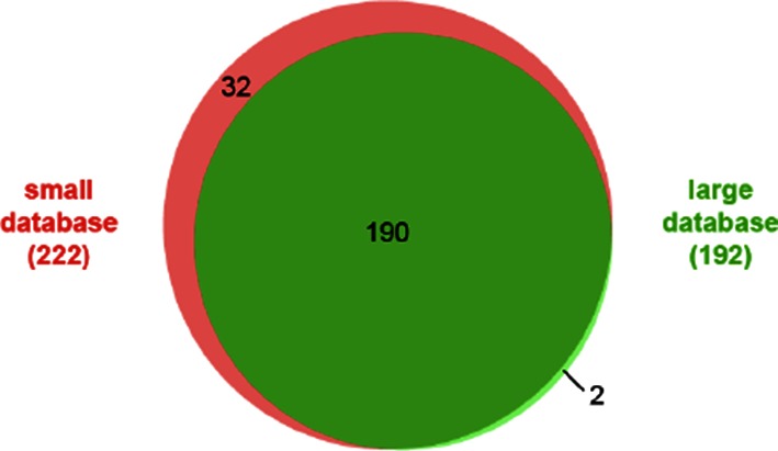
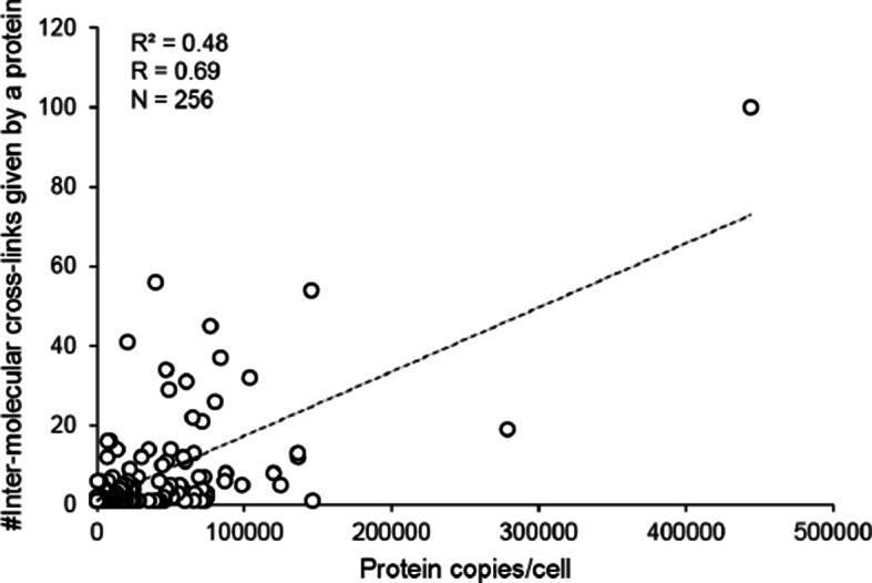
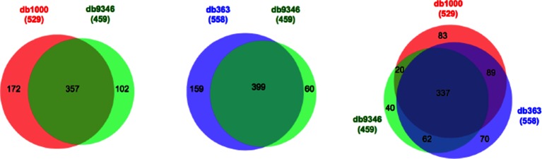
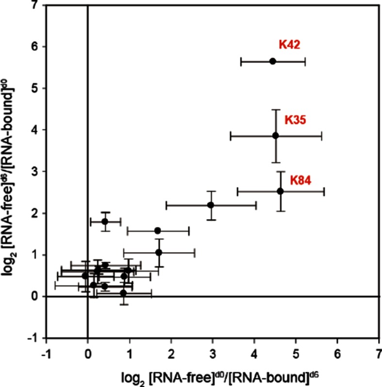
To improve chemical cross-linking of proteins coupled with mass spectrometry (CXMS), we developed a lysine-targeted enrichable cross-linker containing a biotin tag for affinity purification, a chemical cleavage site to separate cross-linked peptides away from biotin after enrichment, and a spacer arm that can be labeled with stable isotopes for quantitation. By locating the flexible proteins on the surface of 70S ribosome, we show that this trifunctional cross-linker is effective at attaining structural information not easily attainable by crystallography and electron microscopy. From a crude Rrp46 immunoprecipitate, it helped identify two direct binding partners of Rrp46 and 15 protein-protein interactions (PPIs) among the co-immunoprecipitated exosome subunits. Applying it to E. coli and C. elegans lysates, we identified 3130 and 893 inter-linked lysine pairs, representing 677 and 121 PPIs. Using a quantitative CXMS workflow we demonstrate that it can reveal changes in the reactivity of lysine residues due to protein-nucleic acid interaction.

Keywords: 70S ribosome; c. elegans; e. coli; biophysics; cross-linking; exosome; mass spectrometry; protein structure; protein-protein interactions; structural biology.


Conflict of interest statement

The author declares that no competing interests exist.

Figures

Figure 1.
Figure 1.. Chemical structures of different designs of Leiker.
The top panel shows four designs of two-piece Leiker with a photo-cleavage site (sulfo-PL, PL, and PEG-PL) or an azobenzene-based cleavage site (AL). Biotin is attached via click chemistry by reacting with bio-aizde. The bottom panel shows two unlabeled (bAL1, bAL2) and deuterium-labeled ([d6]-bAL2) one-piece Leiker molecules. The biotin moiety is colored magenta. DOI: http://dx.doi.org/10.7554/eLife.12509.003
Figure 1—figure supplement 1.
Figure 1—figure supplement 1.. Optimization of protein-to-cross-linker ratio (w/w) for (A) sulfo-PL, (B) AL, (C) bAL1, and (D) bAL2.
DOI: http://dx.doi.org/10.7554/eLife.12509.004
Figure 1—figure supplement 2.
Figure 1—figure supplement 2.. Evaluation of azobenzene-based chemical cleavage.
DOI: http://dx.doi.org/10.7554/eLife.12509.005
Figure 1—figure supplement 3.
Figure 1—figure supplement 3.. The one-piece Leiker (bAL1) outperformed the two-piece Leiker (AL) in the CXMS analysis of a mixture of ten standard proteins.
DOI: http://dx.doi.org/10.7554/eLife.12509.006
Figure 1—figure supplement 4.
Figure 1—figure supplement 4.. Evaluation of the two piece Azo-Leiker (AL).
DOI: http://dx.doi.org/10.7554/eLife.12509.007
Figure 1—figure supplement 5.
Figure 1—figure supplement 5.. bAL1 and bAL2 performed similarly.
DOI: http://dx.doi.org/10.7554/eLife.12509.008
Figure 1—figure supplement 6.
Figure 1—figure supplement 6.. MS1 spectra of (A) [d0]-bAL2 and (B) [d6]-bAL2.
DOI: http://dx.doi.org/10.7554/eLife.12509.009
Figure 2.
Figure 2.. Scheme of the Leiker-based CXMS workflow.
(A) Leiker contains a biotin moiety (magenta), a cleavage site (arrows), and six hydrogen atoms that are accessible to isotope labeling (asterisks). (B) The workflow for purification of Leiker-linked peptides. (C) Three types of Leiker-linked peptides. (D) Leiker-linked peptides generate a reporter ion of 122.06 m/z in HCD, as shown in the spectrum of an inter-linked peptide NYQEAKDAFLGSFLYEYSR-LAKEYEATLEECCAK (+4 charged, MH+ 4433.0553), in which C denotes carbamidomethylated cysteine. DOI: http://dx.doi.org/10.7554/eLife.12509.010
Figure 3.
Figure 3.. Evaluating the performance of Leiker.
(A) Leiker allowed near 100% enrichment of target peptides from a cross-linked ten-protein mixture diluted with increasing amounts of non-cross-linked E. coli lysates. Dark blue, inter-links; light blue, mono-links; green, loop-links; grey, regular peptides not modified by Leiker. (B) Number of cross-link identifications from E. coli lysates treated with Leiker or BS3. Shown in the left and right panels are the identified spectra and peptides, respectively. DOI: http://dx.doi.org/10.7554/eLife.12509.011
Figure 3—figure supplement 1.
Figure 3—figure supplement 1.. Distance distributions of cross-linked lysine pairs in the undiluted ten-protein mixture.
DOI: http://dx.doi.org/10.7554/eLife.12509.014
Figure 4.
Figure 4.. Leiker-based CXMS analyses of large protein assemblies.
(A) Analysis of a purified E. coli 70S ribosome revealed the locations of highly dynamic periphery ribosomal proteins S1, L1, and L7/12 that were refractory to crystallography and cryo-EM analysis. Cross-links to S1, L1, and L7/12 are colored red, blue, and yellow, respectively, and the cross-linked residues on these three proteins are numbered according to the Uniprot sequences. (B) Analysis of a crude immunoprecipitate of the yeast exosome complex. Dashed blue and grey lines denote 50 compatible and 22 incompatible cross-links, respectively, according to the structure of the RNA-bound 11-subunit exosome complex (PDB code: 4IFD). Rrp44, green; Rrp40, orange; Rrp4, violet; Rrp42, gold; other exosome subunits, yellow; RNA, black. Known and candidate exosome regulators revealed by Leiker-cross-links are shown along the periphery and highlighted in green and yellow circles, respectively. (C) Connectivity maps of the ten-subunit exosome core complex based on the inter-molecular cross-links identified in the current IP-CXMS experiments or on previous yeast two-hybrid (Y2H) studies (Stark et al., 2006; Uetz et al., 2000; Oliveira et al., 2002; Luz et al., 2007; Yu et al., 2008). Blue solid lines: experimentally identified putative direct protein-protein interactions; grey dashed lines: theoretical cross-links according to the crystal structure; Cα-Cα distance cutoff ≤30 Å. DOI: http://dx.doi.org/10.7554/eLife.12509.015
Figure 4—figure supplement 1.
Figure 4—figure supplement 1.. Distance distribution of the inter-molecular and intra-molecular cross-links identified in 70S ribosomes.
DOI: http://dx.doi.org/10.7554/eLife.12509.020
Figure 4—figure supplement 2.
Figure 4—figure supplement 2.. Alignment of L9 and L2 from the crystal structure (L9, orange; L2, wheat; PDB code: 2AW4) and their counterparts from the cryo-EM reconstruction (L9, blue; L2, lightblue; PDB code: 5AFI).
DOI: http://dx.doi.org/10.7554/eLife.12509.021
Figure 4—figure supplement 3.
Figure 4—figure supplement 3.. Negative staining of non-cross-linked E. coli 70S ribosome.
DOI: http://dx.doi.org/10.7554/eLife.12509.022
Figure 4—figure supplement 4.
Figure 4—figure supplement 4.. Connectivity maps of cross-links involving (A) S1, (B) L1, (C) L7/12, and (D) L31.
DOI: http://dx.doi.org/10.7554/eLife.12509.023
Figure 4—figure supplement 5.
Figure 4—figure supplement 5.. Silver-stained SDS-PAGE gel of the crude immunoprecipitate of TAP-tagged Rrp46.
DOI: http://dx.doi.org/10.7554/eLife.12509.024
Figure 4—figure supplement 6.
Figure 4—figure supplement 6.. Number of identified inter-linked peptide pairs from decreasing amount of Leiker-cross-linked exosome immunoprecipitate (FDR < 0.05, E-value < 0.01).
After enrichment, 30% (orange) or 60% (blue) of each sample was analyzed by LC-MS/MS. DOI: http://dx.doi.org/10.7554/eLife.12509.025
Figure 5.
Figure 5.. CXMS analyses of E. coli and C. elegans lysates.
(A) The best protein-protein interaction cluster extracted from the Leiker-identified or BS3-identified (Yang et al., 2012) inter-links from E. coli whole-cell lysates. Node size represents the degree of connectivity of the indicated protein in the network. Line width represents the spectral counts of every inter-molecular cross-link. The line color is set to blue when the two peptides of an inter-link are both attributed to unique proteins, to grey if either could be assigned to multiple proteins. All the lines connected to EF-Tu1 are grey because EF-Tu1 differs from EF-Tu2 by only one amino acid. (B) Comparison of the identified inter-links in E. coli whole-cell lysates and ribo-free lysates (5% FDR, E-value < 0.01, spectral count ≥ 3). (C and D) Comparison of the number of Leiker-identified inter-links and that of BS3-identified inter-links (Yang et al., 2012) from C. elegans (C) and E. coli (D) whole-cell lysates (5% FDR, E-value < 0.01, spectral count ≥ 1). DOI: http://dx.doi.org/10.7554/eLife.12509.026
Figure 5—figure supplement 1.
Figure 5—figure supplement 1.. Fractionation of digested, Leiker-treated E. coli lysates.
DOI: http://dx.doi.org/10.7554/eLife.12509.031
Figure 5—figure supplement 2.
Figure 5—figure supplement 2.. Overlap of cross-linked lysine pairs between biological replicates of E. coli lysates (FDR < 0.05, E-value < 0.01, and spectral count ≥ 3).
DOI: http://dx.doi.org/10.7554/eLife.12509.032
Figure 5—figure supplement 3.
Figure 5—figure supplement 3.. Protein-protein interaction networks constructed from the cross-links identified in (A) E. coli and (B) C. elegans.
The labeling scheme is the same as described in Figure 5A except for the node color. For E. coli, node color is set to orange if the protein was only identified in the whole-cell lysates, to yellow only identified in the ribo-free lysates, or to green if identified in both. There are 626 proteins in the E. coli network and 155 proteins in the C. elegans network. DOI: http://dx.doi.org/10.7554/eLife.12509.033
Figure 6.
Figure 6.. Workflow for quantification of cross-linked peptides using pQuant.
For each identified cross-link spectrum, an extracted ion chromatogram (EIC) is constructed for each isotopic peak of the [d0]- and [d6]-labeled precursor. The [d6]/[d0] ratios can be calculated based on the monoisotopic peak, the most intense peak, or the least interfered peak of each isotopic cluster as specified by users. The accuracy of the ratio calculation was evaluated with the confidence score σ (range: 0–1, from the most to the least reliable). If a cross-link have ratios with σ < 0.5, the median of these ratios is assigned to this cross-link. The cross-link ratios of the proteins of interest are normalized to the median ratio of all BSA cross-links. For each cross-link, the median [state1]/[state2] ratio of three independent forward labeling experiments is plotted against the median ratio of three independent reverse labeling experiments. Cross-links that are only present in state1 or state2 due to a dramatic conformational change cannot be quantified as described above because the ratios would be zero or infinite and their σ values would be 1. Therefore, if a cross-link does not have a valid ratio after automatic quantification, the EICs were manually inspected to determine if it was an all-or-none change. DOI: http://dx.doi.org/10.7554/eLife.12509.034
Figure 7.
Figure 7.. Quantitative CXMS analysis of the L7Ae-RNA complex.
(A) Reciprocal labeling of RNA-free (F) and RNA-bound (B) L7Ae with [d0]/[d6]-Leiker. (B) Abundance ratios of mono-links (F/B) in the forward (F[d0]/B[d6]) and the reverse labeling experiment (F[d6]/B[d0]). Each circle represents a mono-linked lysine residue and is colored red if it has a ratio greater than five in both labeling schemes. (C) The three lysine residues affected by RNA binding are highlighted in the structure model (PDB code: 2HVY). The number below each such lysine residue indicates the buried surface area (Å2) upon RNA binding. (D) Extracted ion chromatograms (left) and representative MS1 spectra (right) of a K42 mono-link. DOI: http://dx.doi.org/10.7554/eLife.12509.035
Figure 7—figure supplement 1.
Figure 7—figure supplement 1.. Extracted ion chromatograms (left) and representative MS1 spectra (right) of a mono-linked peptide corresponding to (A) K35 and (B) K84.
DOI: http://dx.doi.org/10.7554/eLife.12509.037
Figure 8.
Figure 8.. Quantitative CXMS analysis of E. coli lysates.
Abundance ratios of (A) inter-linked lysine pairs and (B) mono-linked sites in the forward ([log phase]d0/[stationary phase]d6) and the reverse labeling experiment ([log phase]d6/[stationary phase]d0). DOI: http://dx.doi.org/10.7554/eLife.12509.038
Appendix 1—figure 1.
Appendix 1—figure 1.. Synthesis of sulfo-Photo-cleavable Leiker (sulfo-PL, 8)
DOI: http://dx.doi.org/10.7554/eLife.12509.040
Appendix 1—figure 2.
Appendix 1—figure 2.. Compound 3.
DOI: http://dx.doi.org/10.7554/eLife.12509.041
Appendix 1—figure 3.
Appendix 1—figure 3.. Compound 6.
DOI: http://dx.doi.org/10.7554/eLife.12509.042
Appendix 1—figure 4.
Appendix 1—figure 4.. Compound 7.
DOI: http://dx.doi.org/10.7554/eLife.12509.043
Appendix 1—figure 5.
Appendix 1—figure 5.. Compound 8.
DOI: http://dx.doi.org/10.7554/eLife.12509.044
Appendix 1—figure 6.
Appendix 1—figure 6.. Synthesis of Azo-Leiker (AL, 13).
DOI: http://dx.doi.org/10.7554/eLife.12509.045
Appendix 1—figure 7.
Appendix 1—figure 7.. Compound 11.
DOI: http://dx.doi.org/10.7554/eLife.12509.046
Appendix 1—figure 8.
Appendix 1—figure 8.. Compound 12.
DOI: http://dx.doi.org/10.7554/eLife.12509.047
Appendix 1—figure 9.
Appendix 1—figure 9.. Compound 13.
DOI: http://dx.doi.org/10.7554/eLife.12509.048
Appendix 1—figure 10.
Appendix 1—figure 10.. Synthesis of biotinylated Azo-Leiker 1 (bAL 1, 17).
DOI: http://dx.doi.org/10.7554/eLife.12509.049
Appendix 1—figure 11.
Appendix 1—figure 11.. Compound 15.
DOI: http://dx.doi.org/10.7554/eLife.12509.050
Appendix 1—figure 12.
Appendix 1—figure 12.. Compound 16.
DOI: http://dx.doi.org/10.7554/eLife.12509.051
Appendix 1—figure 13.
Appendix 1—figure 13.. Compound 17.
DOI: http://dx.doi.org/10.7554/eLife.12509.052
Appendix 1—figure 14.
Appendix 1—figure 14.. Synthesis of biotinylated Azo-Leiker 2 (bAL 2, 27).
DOI: http://dx.doi.org/10.7554/eLife.12509.053
Appendix 1—figure 15.
Appendix 1—figure 15.. Compound 20.
DOI: http://dx.doi.org/10.7554/eLife.12509.054
Appendix 1—figure 16.
Appendix 1—figure 16.. Compound 21.
DOI: http://dx.doi.org/10.7554/eLife.12509.055
Appendix 1—figure 17.
Appendix 1—figure 17.. Compound 22.
DOI: http://dx.doi.org/10.7554/eLife.12509.056
Appendix 1—figure 18.
Appendix 1—figure 18.. Compound 23.
DOI: http://dx.doi.org/10.7554/eLife.12509.057
Appendix 1—figure 19.
Appendix 1—figure 19.. Compound 25.
DOI: http://dx.doi.org/10.7554/eLife.12509.058
Appendix 1—figure 20.
Appendix 1—figure 20.. Compound 26.
DOI: http://dx.doi.org/10.7554/eLife.12509.059
Appendix 1—figure 21.
Appendix 1—figure 21.. Compound 27.
DOI: http://dx.doi.org/10.7554/eLife.12509.060
Appendix 1—figure 22.
Appendix 1—figure 22.. Synthesis of d6-biotinylated Azo-Leiker 2 (bAL 2, 31).
DOI: http://dx.doi.org/10.7554/eLife.12509.061
Appendix 1—figure 23.
Appendix 1—figure 23.. Compound 29.
DOI: http://dx.doi.org/10.7554/eLife.12509.062
Appendix 1—figure 24.
Appendix 1—figure 24.. Compound 30.
DOI: http://dx.doi.org/10.7554/eLife.12509.063
Appendix 1—figure 25.
Appendix 1—figure 25.. Compound 31.
DOI: http://dx.doi.org/10.7554/eLife.12509.064
Appendix 1—figure 26.
Appendix 1—figure 26.. 1H NMR spectra of compound 6.
DOI: http://dx.doi.org/10.7554/eLife.12509.065
Appendix 1—figure 27.
Appendix 1—figure 27.. 13C NMR spectra of compound 6.
DOI: http://dx.doi.org/10.7554/eLife.12509.066
Appendix 1—figure 28.
Appendix 1—figure 28.. 1H NMR spectra of sulfo-PL.
DOI: http://dx.doi.org/10.7554/eLife.12509.067
Appendix 1—figure 29.
Appendix 1—figure 29.. 13C NMR spectra of sulfo-PL.
DOI: http://dx.doi.org/10.7554/eLife.12509.068
Appendix 1—figure 30.
Appendix 1—figure 30.. 1H NMR spectra of compound 11.
DOI: http://dx.doi.org/10.7554/eLife.12509.069
Appendix 1—figure 31.
Appendix 1—figure 31.. 13C NMR spectra of compound 11.
DOI: http://dx.doi.org/10.7554/eLife.12509.070
Appendix 1—figure 32.
Appendix 1—figure 32.. 1H NMR spectra of AL.
DOI: http://dx.doi.org/10.7554/eLife.12509.071
Appendix 1—figure 33.
Appendix 1—figure 33.. 13C NMR spectra of AL.
DOI: http://dx.doi.org/10.7554/eLife.12509.072
Appendix 1—figure 34.
Appendix 1—figure 34.. 1H NMR spectra of compound 15.
DOI: http://dx.doi.org/10.7554/eLife.12509.073
Appendix 1—figure 35.
Appendix 1—figure 35.. 13C NMR spectra of compound 15.
DOI: http://dx.doi.org/10.7554/eLife.12509.074
Appendix 1—figure 36.
Appendix 1—figure 36.. 1H NMR spectra of bAL 1.
DOI: http://dx.doi.org/10.7554/eLife.12509.075
Appendix 1—figure 37.
Appendix 1—figure 37.. 13C NMR spectra of bAL 1.
DOI: http://dx.doi.org/10.7554/eLife.12509.076
Appendix 1—figure 38.
Appendix 1—figure 38.. 1H NMR spectra of compound 20.
DOI: http://dx.doi.org/10.7554/eLife.12509.077
Appendix 1—figure 39.
Appendix 1—figure 39.. 13C NMR spectra of compound 20.
DOI: http://dx.doi.org/10.7554/eLife.12509.078
Appendix 1—figure 40.
Appendix 1—figure 40.. 1H NMR spectra of compound 21.
DOI: http://dx.doi.org/10.7554/eLife.12509.079
Appendix 1—figure 41.
Appendix 1—figure 41.. 13C NMR spectra of compound 21.
DOI: http://dx.doi.org/10.7554/eLife.12509.080
Appendix 1—figure 42.
Appendix 1—figure 42.. 1H NMR spectra of compound 22.
DOI: http://dx.doi.org/10.7554/eLife.12509.081
Appendix 1—figure 43.
Appendix 1—figure 43.. 13C NMR spectra of compound 22.
DOI: http://dx.doi.org/10.7554/eLife.12509.082
Appendix 1—figure 44.
Appendix 1—figure 44.. 1H NMR spectra of compound 23.
DOI: http://dx.doi.org/10.7554/eLife.12509.083
Appendix 1—figure 45.
Appendix 1—figure 45.. 13C NMR spectra of compound 23.
DOI: http://dx.doi.org/10.7554/eLife.12509.084
Appendix 1—figure 46.
Appendix 1—figure 46.. 1H NMR spectra of compound 25.
DOI: http://dx.doi.org/10.7554/eLife.12509.085
Appendix 1—figure 47.
Appendix 1—figure 47.. 13C NMR spectra of compound 25.
DOI: http://dx.doi.org/10.7554/eLife.12509.086
Appendix 1—figure 48.
Appendix 1—figure 48.. 1H NMR spectra of bAL 2.
DOI: http://dx.doi.org/10.7554/eLife.12509.087
Appendix 1—figure 49.
Appendix 1—figure 49.. 13C NMR spectra of bAL 2.
DOI: http://dx.doi.org/10.7554/eLife.12509.088
Appendix 1—figure 50.
Appendix 1—figure 50.. 1H NMR spectra of compound 29.
DOI: http://dx.doi.org/10.7554/eLife.12509.089
Appendix 1—figure 51.
Appendix 1—figure 51.. 13C NMR spectra of compound 29.
DOI: http://dx.doi.org/10.7554/eLife.12509.090
Appendix 1—figure 52.
Appendix 1—figure 52.. 1H NMR spectra of d6-bAL 2.
DOI: http://dx.doi.org/10.7554/eLife.12509.091
Appendix 1—figure 53.
Appendix 1—figure 53.. 13C NMR spectra of d6-bAL 2.
DOI: http://dx.doi.org/10.7554/eLife.12509.092
Author response image 1.
Author response image 1.. Overlap of inter-links between subunits of the exosome core complex.
DOI: http://dx.doi.org/10.7554/eLife.12509.094
Author response image 2.
Author response image 2.. Intensity distributions of the reporter ion of m/z 122.0606 for different types of peptides.
In each category, the FDR of peptide-spectrum match (PSM) is 5%. The data came from the ten standard proteins. DOI: http://dx.doi.org/10.7554/eLife.12509.095
Author response image 3.
Author response image 3.. Silver-stained SDS-PAGE of the crude immunoprecipitate of TAP-tagged Rrp46 (left) and the SDS-PAGE of the purified exosome sample of Shi et al. (right).
DOI: http://dx.doi.org/10.7554/eLife.12509.096
Author response image 4.
Author response image 4.. Overlap between two CXMS experiments using BS3 or Leiker on the same 10-protein mix.
DOI: http://dx.doi.org/10.7554/eLife.12509.097
Author response image 5.
Author response image 5.. Comparison of identified cross-links using a small database (62 proteins) and a large database (562 proteins).
Filtering criteria: FDR < 5%, E-value < 0.00001, and spectral count ≥ 5. DOI: http://dx.doi.org/10.7554/eLife.12509.098
Author response image 6.
Author response image 6.. The number of inter-molecular cross-links observed for a protein in E. coli whole-cell lysates is positively correlated with the abundance of this protein.
R, Pearson correlation coefficients; N, number of values. DOI: http://dx.doi.org/10.7554/eLife.12509.099
Author response image 7.
Author response image 7.. Comparison of identified cross-links using databases of different size.
db9346, containing all the proteins identified in the sample; db1000, containing the top 1000 abundant proteins; db363, containing all the proteins for which intra-molecular cross-links had been identified. Filtering criteria: FDR < 5%, E-value < 0.01, and spectral count ≥ 3. DOI: http://dx.doi.org/10.7554/eLife.12509.100
Author response image 8.
Author response image 8.. Abundance ratios of mono-links (F/B) in the forward (F[d0]/B[d6]) and the reverse labeling experiment (F[d6]/B[d0]).
Mean ± SEM. DOI: http://dx.doi.org/10.7554/eLife.12509.102

References

    1. Araki Y, Takahashi S, Kobayashi T, Kajiho H, Hoshino S, Katada T. Ski7p G protein interacts with the exosome and the Ski complex for 3'-to-5' mRNA decay in yeast. The EMBO Journal. 2001;20:4684–4693. doi: 10.1093/emboj/20.17.4684. - DOI - PMC - PubMed
    1. Bader GD, Hogue CWV. An automated method for finding molecular complexes in large protein interaction networks. BMC Bioinformatics. 2003;4:2. doi: 10.1186/1471-2105-4-2. - DOI - PMC - PubMed
    1. Balandina A, Claret L, Hengge-Aronis R, Rouviere-Yaniv J. The Escherichia coli histone-like protein HU regulates rpoS translation. Molecular Microbiology. 2001;39:1069–1079. doi: 10.1046/j.1365-2958.2001.02305.x. - DOI - PubMed
    1. Bohn S, Beck F, Sakata E, Walzthoeni T, Beck M, Aebersold R, Förster F, Baumeister W, Nickell S. Structure of the 26S proteasome from Schizosaccharomyces pombe at subnanometer resolution. Proceedings of the National Academy of Sciences of the United States of America. 2010;107:20992–20997. doi: 10.1073/pnas.1015530107. - DOI - PMC - PubMed
    1. Brandt F, Etchells SA, Ortiz JO, Elcock AH, Hartl FU, Baumeister W. The native 3D organization of bacterial polysomes. Cell. 2009;136:261–271. doi: 10.1016/j.cell.2008.11.016. - DOI - PubMed

Publication types

MeSH terms

Substances