Review
. 2016 Jul;1863(7 Pt B):1717-27.
doi: 10.1016/j.bbamcr.2016.03.003. Epub 2016 Mar 4.

High throughput physiological screening of iPSC-derived cardiomyocytes for drug development

Affiliations
Review

High throughput physiological screening of iPSC-derived cardiomyocytes for drug development

Juan C Del Álamo et al. Biochim Biophys Acta. 2016 Jul.

Abstract

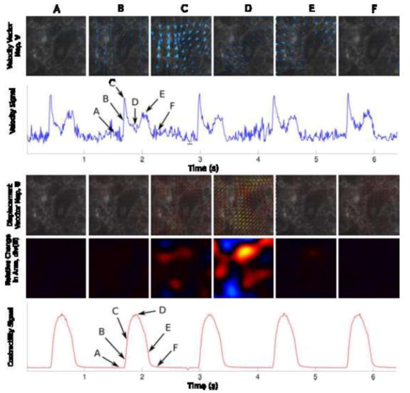
Cardiac drug discovery is hampered by the reliance on non-human animal and cellular models with inadequate throughput and physiological fidelity to accurately identify new targets and test novel therapeutic strategies. Similarly, adverse drug effects on the heart are challenging to model, contributing to costly failure of drugs during development and even after market launch. Human induced pluripotent stem cell derived cardiac tissue represents a potentially powerful means to model aspects of heart physiology relevant to disease and adverse drug effects, providing both the human context and throughput needed to improve the efficiency of drug development. Here we review emerging technologies for high throughput measurements of cardiomyocyte physiology, and comment on the promises and challenges of using iPSC-derived cardiomyocytes to model disease and introduce the human context into early stages of drug discovery. This article is part of a Special Issue entitled: Cardiomyocyte biology: Integration of Developmental and Environmental Cues in the Heart edited by Marcus Schaub and Hughes Abriel.

Keywords: Automated microscopy; Cardiomyocyte; Drug discovery; Heart; High content screening; Particle image velocimetry; Physiology.


Figures

Figure 1
Figure 1. Matrigel micropatterns on traction-sensitive polyacrylamide hydrogel devices constrain hPSC-CMs to controllable shapes and engineer their mechanical output
A) Human iPSC-cardiomyocytes were cultured on micropatterns to induce aspect ratios of 1:1–7:1 and areas of 2,000 μm2. Lifeact-labeled actin in myofibrils in live iPSC-cardiomyocytes show sarcomeric organization. B) Myofibril alignment leads to higher contractile forces in single human iPSC-cardiomyocytes. Σ|Fc| of engineered hPSC-CMs increases with aspect ratio of the cardiomyocytes on the micropatterned surfaes. C) iPSC-cardiomyocytes labeled with di-8-ANEPPS to reveal T-tubules. (Left) iPSC-cardiomyocyte (7:1) (Scale bar, 25 μm.). (Center) Unpatterned iPSC-cardiomyocyte on a 10-kPa hydrogel shown in both Top and Bottom (Scale bars, 50 μm.). (Right) Isolated adult mouse ventricular cardiomyocyte (Scale bar, 25 μm.).
Figure 2
Figure 2. Measurement of iPSC-cardiomyocyte contractility from deformation maps
Top Row) Velocity maps shown at 6 different timeframes (A through F) corresponding to relevant events in the contraction cycle: Relaxed State, Beginning of the Contraction, Maximum Contraction Rate, Maximum Contraction, Maximum Relaxation Rate, and Relaxed State again. Second Row) The Velocity Signal is obtained by taking the average of the magnitude of the each instantaneous velocity maps. The frame selection was done automatically. Selected reference frames (frames with least motion) are underlined in black. Third Row) Displacement Vector Maps are computed using the individual timepoints relative to the reference frames. The 6 images represent the same maps (A through F) shown in the First Row. Fourth Row) Within the displacement vector maps, the relative change in area (magnitude in color) is computed by direct application of Gauss’ theorem. Fifth Row) A contractility signal is obtained by taking the average of the magnitude of the relative change in area.
Figure 3
Figure 3. Modeling lipotoxicity in an iPSC-CM model
A) iPSC-CMs cultured in low fat media (supplemented with 25μM linoleic acid-oleic acid-albumin; L-O-BSA – Sigma, L9655) for one week have very few adiposomes. B) Culturing iPSC-CMs with a “high fat diet” (HFD - 250μM L-O-BSA) results in increased number of adiposomes per cell. C–E) This phenotype is recapitulated by culture in low fat media at hypoxia (2% O2 for 48hrs) (C) or by siRNA-mediated PNPLA2 knockdown (D), and is exacerbated by the combination of hypoxia and HFD (E). F) Quantification of the nile red-stained adiposomes (see A inset for example) using CellProfiler software

Similar articles

Cited by

References

    1. Balinsky BI. Experiments on total extirpation of the whole endoderm in Triton embryos. C.r Acad Sci URSS. 1939;23:196–198.
    1. Chuang HH, Tseng MP. An experimental analysis of the determination and differentiation of the mesodermal structures of neurula in urodeles. Scientia Sinica. 1957;6:669–708. - PubMed
    1. Jacobson AG. Influences of ectoderm and endoderm on heart differentiation in the newt. Developmental Biology. 1960;2:138–154. - PubMed
    1. Jacobson AG. Heart determination in the newt. Journal of Experimental Zoology. 1961;146:139–152. - PubMed
    1. Nieuwkoop PD. Experimental investigations on the origin and determination of the germ cells, and on the development of the lateral plates and germ ridges in Urodeles. Archs Neerl Zool. 1947;8:1–205.

Publication types

MeSH terms

Substances