. 2016 Apr 15;143(8):1318-27.
doi: 10.1242/dev.129122. Epub 2016 Mar 7.

Developmental origin of lung macrophage diversity

Affiliations

Developmental origin of lung macrophage diversity

Serena Y S Tan et al. Development. .

Abstract

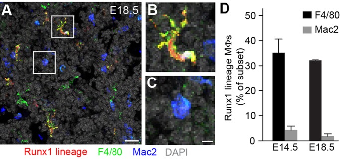
Macrophages are specialized phagocytic cells, present in all tissues, which engulf and digest pathogens, infected and dying cells, and debris, and can recruit and regulate other immune cells and the inflammatory response and aid in tissue repair. Macrophage subpopulations play distinct roles in these processes and in disease, and are typically recognized by differences in marker expression, immune function, or tissue of residency. Although macrophage subpopulations in the brain have been found to have distinct developmental origins, the extent to which development contributes to macrophage diversity between tissues and within tissues is not well understood. Here, we investigate the development and maintenance of mouse lung macrophages by marker expression patterns, genetic lineage tracing and parabiosis. We show that macrophages populate the lung in three developmental waves, each giving rise to a distinct lineage. These lineages express different markers, reside in different locations, renew in different ways, and show little or no interconversion. Thus, development contributes significantly to lung macrophage diversity and targets each lineage to a different anatomical domain.

Keywords: Lineage tracing; Lung development; Lung macrophage; Parabiosis; Phagocytosis.


Conflict of interest statement

Competing interests

The authors declare no competing or financial interests.

Figures

Fig. 1.
Fig. 1.
Marker expression and morphology of mid-embryonic lung macrophages. (A-C) Lung sections from E15.5 c-fms-EGFP mice immunostained as indicated for macrophage markers F4/80 and Mac2, and for E-cadherin (gray) to show airway epithelium. All lung macrophages express c-fms-EGFP (green) and major subsets express F4/80 (A) or Mac2 (B). Co-staining for F4/80 and Mac2 (C) show that all c-fms+ macrophages express either high F4/80 or Mac2. (D) E15.5 wild-type (C57BL/6J) lung co-stained for F4/80 and Mac2. F4/80 and Mac2 label complementary macrophage subsets. (E,F) Close-ups of the boxed regions in D. F4/80+ macrophages are more irregularly shaped with pseudopodia (E), whereas Mac2+ macrophages are more regular and elliptical (F). DAPI, nuclear stain. Scale bars: 20 μm (A-D), 5 μm (E,F).
Fig. 2.
Fig. 2.
Dynamics of F4/80 and Mac2 macrophage subpopulations in the developing lung. (A-D) Sections through wild-type lungs of the indicated embryonic ages immunostained to show F4/80+ (green) and Mac2+ (blue, arrowheads) macrophages. The two subpopulations and their kinetics are consistent with the F4/80-high and F4/80-low macrophage subpopulations previously described in the embryo (Schulz et al., 2012), and indeed low levels of F4/80 can be detected in some Mac2 macrophages especially after E15.5 (Table S1). Scale bars: 20 μm. (E) Quantification of abundance of F4/80+ and Mac2+ macrophages throughout embryonic lung development. F4/80 macrophages dominate early in lung development and Mac2 macrophages dominate after E15.5. Values shown are mean±s.e.m. for n=750-3570 macrophages scored in two or three lungs per time point. Values at early time points obscured by the x-axis are: E9.5 (0 per lung, F4/80 macrophages; 0 per lung, Mac2 macrophages), E10.5 (4±1; 0), E11.5 (39±5; 0), E12.5 (2600±81; 67±21). Arrows indicate the earliest developmental ages that F4/80 (green arrow) and Mac2 macrophages (blue arrow) were detected in the lung. Note that most of the error bars are very small and obscured by the data point symbols. Mɸs, macrophages.
Fig. 3.
Fig. 3.
Lineage tracing of yolk sac precursors in embryonic lung. (A-D) Section through E18.5 lung of a Runx1-CreER>TdTomato mouse (lineage trace in red), induced with tamoxifen at E6.75 to label yolk sac precursors, and immunostained to show F4/80 (green) and Mac2 (blue) macrophages. Lineage label marks F4/80 macrophages (A,B; yellow cells) but few Mac2 macrophages (A,C; blue cells). At this age, many Mac2 macrophages express low, variable levels of F4/80. Scale bars: 20 μm (A), 5 μm (B,C). (D) Quantification showing fraction of each macrophage subpopulation that expresses the Runx1 lineage label at E14.5 and E18.5. Values shown are mean+s.e.m.; n=210-1390 macrophages scored of each type in two lungs per time point. Mɸs, macrophages.
Fig. 4.
Fig. 4.
Lineage trace of yolk sac precursors in the adult lung. (A-E) Adult (P64) lungs from Runx1-CreER>TdTomato lineage trace induced at E6.75 as in Fig. 3 and immunostained for MHCII (green; A-C) or Mac2 (green; D,E) and PECAM (blood vessels, gray) and counterstained with DAPI (blue). Note that lineage-labeled interstitial macrophages express MHCII macrophage marker (yellow cells in A and close-up of peripheral region shown in C) but not Mac2 macrophage marker (red cells in D and perivascular close-up in E) and localize to the periphery along mesothelium (meso; A,C) and large blood vessels (D,E). The macrophages that are not lineage labeled are MHCII+ interstitial macrophages (green cells in A and central close-up in B), located in the more central parenchyma further from the mesothelium, and Mac2+ alveolar macrophages (green cells in D,E). Scale bars: 100 μm (A,D), 20 μm (C,E).
Fig. 5.
Fig. 5.
Gene expression changes in interstitial macrophages after birth. (A-D) Sections of c-fms-EGFP mouse lungs at the indicated postnatal ages immunostained for either F4/80 (A,B) or MHCII (C,D) and wheat germ agglutinin (WGA; A) to show epithelium or PECAM (B-D) to show blood vessels. Because c-fms is a robust and specific macrophage marker, whereas F4/80 is not always macrophage restricted, only c-fms-EGFP+ cells were considered macrophages and the identity of the F4/80+ c-fms cells was not determined. Interstitial macrophages (encircled) were distinguished from alveolar macrophages by their location within the walls separating alveolar lumens, and their distinctive dendritic-like morphology. Arrowheads indicate examples of type II alveolar epithelial cells, which express MHCII (Debbabi, 2005) but not c-fms (C,D). Scale bar: 20 μm. (E) Quantification of postnatal changes in abundance of F4/80+ and MHCII+ interstitial macrophages. Values shown are mean±s.e.m. of n=100-210 interstitial macrophages scored in two or three lungs at each time point. Note diminution in F4/80+ population and concomitant increase in MHCII+ population in the first 3 weeks of life. A small transient postnatal population of interstitial macrophages that are F4/80 and MHCII is not depicted in the graph. (F) Quantification of postnatal changes in abundance of F4/80+ and F4/80Cre-lineage-positive interstitial macrophages. Note the spike at P4 in F4/80+, F4/80Cre-lineage-negative interstitial macrophages (blue line). Values shown are mean±s.e.m. of n=90-170 interstitial macrophages scored in two or three lungs at each time point; many of the error bars are small and obscured by the data point symbols. Mɸs, macrophages.
Fig. 6.
Fig. 6.
Replenishment of interstitial but not alveolar macrophages during parabiosis. (A) Lung of a host (unlabeled) C57BL/6J P98 adult mouse after parabiosis for 1 month with a litter mate donor C57BL/6 CAG-EGFP mouse that expresses EGFP (green) ubiquitously. Lung was immunostained for CD68+ (all macrophages) and WGA (epithelium) and counterstained with DAPI. Note that some interstitial CD68+ macrophages are GFP labeled and hence donor derived (encircled green and white in the merge image), whereas the rest of the interstitial macrophages (encircled white in the merge image) and all alveolar macrophages (encircled red in the merge image) are host derived. Scale bar: 40 μm. (B) Quantification of host interstitial and alveolar macrophages that are donor derived (GFP+) following parabiosis. Note replenishment of interstitial but not alveolar macrophages. Values shown are from n=130-160 donor macrophages scored in one animal per time point. Mɸs, macrophages.
Fig. 7.
Fig. 7.
Mac2 macrophages populate the alveoli in the first week after birth. (A,B) Sections of lungs from wild-type mice at the indicated postnatal ages that were immunostained for macrophage markers F4/80 and Mac2, and WGA (epithelium). At birth (A), nearly all Mac2 macrophages are interstitial (arrowheads) but by P6 (B) nearly all Mac2 macrophages are within alveoli (circles), with only a few still interstitial (arrowhead). F4/80 macrophages (green) remain interstitial: inspection of serial optical sections confirmed that the occasional F4/80 macrophage that in a single optical section can appear lumenal (arrows) are actually interstitial. At the stages shown, Mac2 macrophages express low, variable levels of F4/80 as well as WGA. Scale bar: 40 μm. (C) Quantification of Mac2 macrophage localization one day before birth (E18.5) and during first week after birth. Values shown are mean±s.e.m. for n=140-450 macrophages scored in three sections of each lung at each time point; some error bars are very small and obscured by the data point symbols.
Fig. 8.
Fig. 8.
Model of developmental origin of lung macrophage diversity. Timeline (top) and schematics (middle) of lungs at E12, E16, and in the adult illustrating the major developmental events and global localization of the three lung macrophage lineages shown in green, blue and red. Schematics at the bottom are close-ups showing interstitial and lumenal localization of the macrophage lineages, with airway epithelial cells indicated in white, the airway lumen in gray and the interstitium stippled. F4/80 embryonic macrophages (green) arrive from yolk sac in the earliest wave beginning at E10.5. Two days later, Mac2 embryonic macrophages (blue) begin arriving, most likely from the fetal liver (Guilliams et al., 2013). During the first week of postnatal life, Mac2 macrophages enter the lumen to become alveolar macrophages, which self-renew. F4/80 embryonic macrophages persist at specific locations (peripheral and perivascular) to become the ‘primitive’ interstitial macrophages, whereas those in the parenchyma are replaced by a new population of ‘definitive’ interstitial macrophages (red) from the circulation, presumably of bone marrow origin. Both F4/80 lineages turn off F4/80 and turn on MHCII during the first 3 weeks of postnatal life (not shown). Meso, mesothelium; MΦ, macrophage.

References

    1. Aguzzi A., Barres B. A. and Bennett M. L. (2013). Microglia: scapegoat, saboteur, or something else? Science 339, 156-161. 10.1126/science.1227901 - DOI - PMC - PubMed
    1. Bedoret D., Wallemacq H., Marichal T., Desmet C., Quesada Calvo F., Henry E., Closset R., Dewals B., Thielen C., Gustin P. et al. (2009). Lung interstitial macrophages alter dendritic cell functions to prevent airway allergy in mice. J. Clin. Invest. 119, 3723-3738. 10.1172/JCI39717 - DOI - PMC - PubMed
    1. Blussé van Oud Alblas A., van der Linden-Schrever B. and van Furth R. (1981). Origin and kinetics of pulmonary macrophages during an inflammatory reaction induced by intravenous administration of heat-killed bacillus Calmette-Guerin. J. Exp. Med. 154, 235-252. 10.1084/jem.154.2.235 - DOI - PMC - PubMed
    1. Bowden D. H. and Adamson I. Y. (1972). The pulmonary interstitial cell as immediate precursor of the alveolar macrophage. Am. J. Pathol. 68, 521-537. - PMC - PubMed
    1. Bowden D. H., Adamson I. Y., Grantham W. G. and Wyatt J. P. (1969). Origin of the lung macrophage. Evidence derived from radiation injury. Arch. Pathol. 88, 540-546. - PubMed

Publication types

Substances