. 2016 Jun;65(6):1660-71.
doi: 10.2337/db15-1127. Epub 2016 Mar 7.

Inhibition of DYRK1A Stimulates Human β-Cell Proliferation

Affiliations

Inhibition of DYRK1A Stimulates Human β-Cell Proliferation

Ercument Dirice et al. Diabetes. 2016 Jun.

Abstract

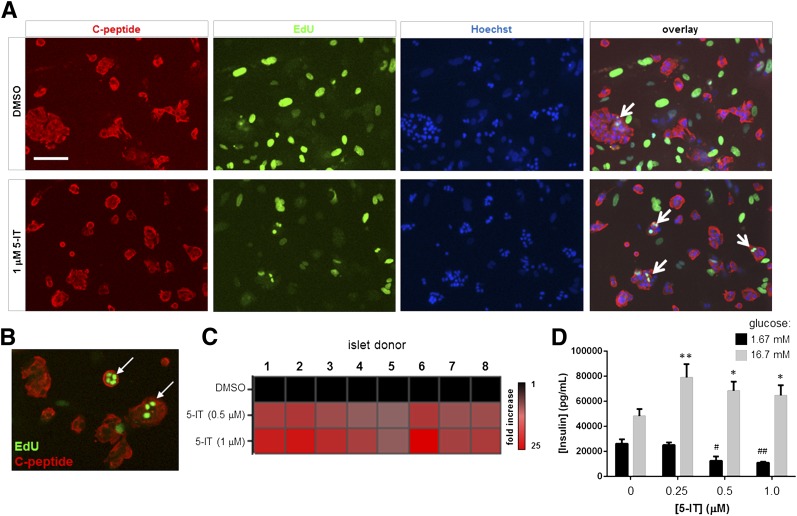
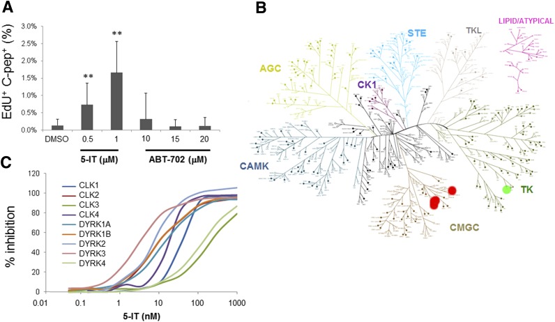
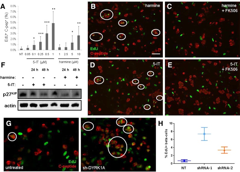
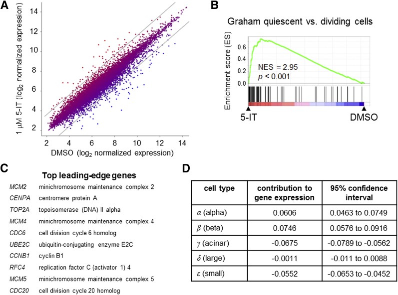
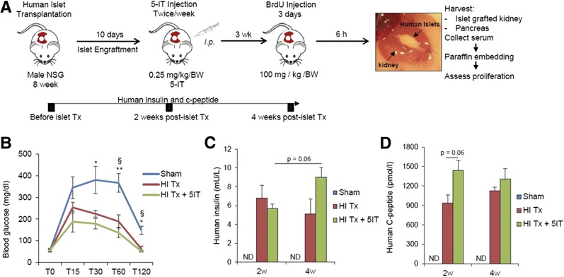
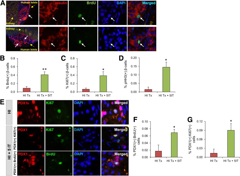
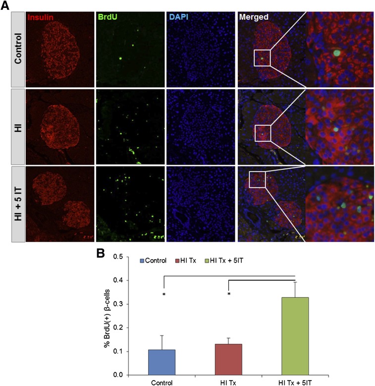
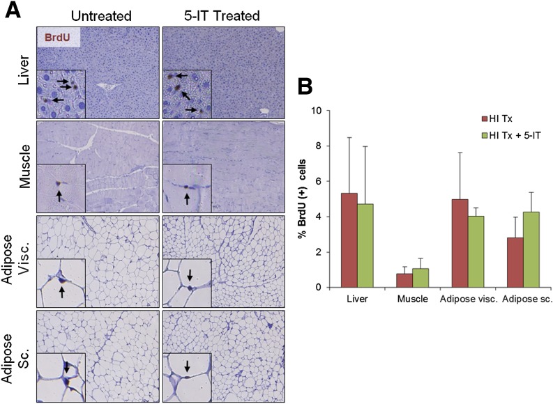
Restoring functional β-cell mass is an important therapeutic goal for both type 1 and type 2 diabetes (1). While proliferation of existing β-cells is the primary means of β-cell replacement in rodents (2), it is unclear whether a similar principle applies to humans, as human β-cells are remarkably resistant to stimulation of division (3,4). Here, we show that 5-iodotubercidin (5-IT), an annotated adenosine kinase inhibitor previously reported to increase proliferation in rodent and porcine islets (5), strongly and selectively increases human β-cell proliferation in vitro and in vivo. Remarkably, 5-IT also increased glucose-dependent insulin secretion after prolonged treatment. Kinome profiling revealed 5-IT to be a potent and selective inhibitor of the dual-specificity tyrosine phosphorylation-regulated kinase (DYRK) and cell division cycle-like kinase families. Induction of β-cell proliferation by either 5-IT or harmine, another natural product DYRK1A inhibitor, was suppressed by coincubation with the calcineurin inhibitor FK506, suggesting involvement of DYRK1A and nuclear factor of activated T cells signaling. Gene expression profiling in whole islets treated with 5-IT revealed induction of proliferation- and cell cycle-related genes, suggesting that true proliferation is induced by 5-IT. Furthermore, 5-IT promotes β-cell proliferation in human islets grafted under the kidney capsule of NOD-scid IL2Rg(null) mice. These results point to inhibition of DYRK1A as a therapeutic strategy to increase human β-cell proliferation.


Figures

Figure 1
Figure 1
Induction of human β-cell proliferation by 5-IT. A: Images showing insulin immunofluorescence and EdU incorporation in dissociated islets treated for 6 days with 1 μmol/L 5-IT. Representative EdU-positive β-cells are indicated with a white arrow. Scale bar, 100 μm. B: Higher-magnification image of EdU-positive β-cells. C: Heat map of EdU-positive β-cells in eight individual islet donors. Donor information is provided in Supplementary Fig. 2. D: GSIS after treatment of dissociated islets for 12 days with the indicated concentration of 5-IT. Cells were challenged with 1.67 mmol/L (black bars) or 16.7 mmol/L glucose (gray bars) for 1 h. Data represent the mean ± SD of four independent wells. *P < 0.01 and **P < 0.001, compared with DMSO treatment (high glucose), two-tailed Student t test. #P < 0.001, compared with DMSO treatment; ##P < 10−4, compared with DMSO treatment (low glucose), two-tailed Student t test.
Figure 2
Figure 2
Inhibition of DYRK and CLK kinase families by 5-IT. A: Comparison of the effects of 6-day treatment with 5-IT and the structurally unrelated adenosine kinase inhibitor ABT-702. The percent of β-cells positive for EdU was measured and calculated. Data represent the mean ± SD of four independent wells and nine fields of view per well. **P < 0.01, compared with DMSO treatment, two-tailed Student t test. B: Kinome profiling of 253 human kinases. Tested kinases are indicated by closed circles, kinases inhibited by 5-IT are indicated in red, and the kinase inhibited by ABT-702 is indicated in green. C: Dose-dependent inhibition of indicated DYRK and CLK by 5-IT.
Figure 3
Figure 3
DYRK1A inhibitor harmine also induces β-cell proliferation. A: Effect of 6-day treatment with indicated concentrations of 5-IT or harmine. Data represent the mean ± SD of four independent wells and nine fields of view per well. *P < 0.05, **P < 0.01, and ***P < 0.001, compared with DMSO treatment, two-tailed Student t test. C-pep, C-peptide. B: Fluorescent image of dissociated islets treated for 6 days with 10 μmol/L harmine. Scale bar, 100 μm. EdU-positive β-cells are circled in white. C: Fluorescent image of dissociated islets treated with 10 μmol/L harmine and 1 μmol/L FK506. D: Fluorescent image of dissociated islets treated for 6 days with 0.5 μmol/L 5-IT. E: Fluorescent image of dissociated islets treated with 0.5 μmol/L 5-IT and 1 μmol/L FK506. F: Western blot of p27KIP phosphorylation (S10) in INS-1E cells in response to 24- or 48-h treatment with 10 μmol/L harmine or 0.5 μmol/L 5-IT. Actin was included as a loading control. G: Fluorescent image of dissociated islets untreated (left) or infected with adenovirus expressing sh-DYRK1A (right). H: Quantification of EdU-positive β-cells in untreated islets or islets in which DYRK1A has been knocked down by two different constructs. Data represent 12 independent wells per condition, imaged at nine sites per well. Error bars represent the 95% CI of the mean. NT, untreated.
Figure 4
Figure 4
Gene expression profiling of 5-IT–treated islets consistent with increased β-cell proliferation. A: Scatter plot of gene expression results of intact islets treated for 6 days with DMSO or 1 μmol/L 5-IT, expressed as the geometric average across three donors. Gray lines show the threshold for genes with threefold increase or decrease in gene expression. B: GSEA results reveal a number of sets associated with cell division. Representative gene set analysis shown. Statistical analysis performed as previously described (23,24). C: Top leading-edge genes contributing to GSEA. D: Estimated changes in cell mass (α, β, acinar, large duct, small duct, and uncharacterized) after treatment with 5-IT, determined by five-dimensional linear regression.
Figure 5
Figure 5
Study design and functionality of human islets grafted under the kidney capsule of NSG mice. A: Experimental strategy showing human islet transplantation under the kidney capsule of male NSG mice followed by either vehicle or intraperitoneal 5-IT (0.25 mg/kg/BW) injection twice a week. BrdU (100 mg/kg/BW) was injected 3 weeks posttransfer for 3 days, and 6 h after the last injection, pancreata were harvested for immunohistochemical analyses. Image shows human islets under the kidney at the end of the follow-up. B: Glucose tolerance test performed 4 weeks after human islet transplantation on sham-operated, vehicle (HI Tx), or 5-IT–treated (HI Tx + 5-IT) mice grafted with human islets (n = 3–4). Glucose was measured at the indicated time (minutes) of the tolerance test. Human insulin (C) and human C-peptide (D) ELISA results assayed from three different groups 2 and 4 weeks post–human islet transplantation. Representative experiment from n = 4. Error bars indicate ±SEM. P values were determined by unpaired Student t test. *P < 0.05; **P < 0.01. §Sham vs. HI Tx; *sham vs. HI Tx + 5-IT. ND, not detectable.
Figure 6
Figure 6
5-IT treatment promotes human β-cell proliferation in vivo. A: Immunohistochemistry of vehicle or 5-IT–treated kidney sections grafted with human islets (10×) showing BrdU (green), insulin (red), and DAPI (blue). Yellow arrows define the kidney and human islet grafts. White arrows mark proliferating β-cells. Magnified images (×40) show each immunofluorescent staining separately (indicating nuclear gap for replicating β-cell surrounded by insulin) for the proliferating β-cell. Representative experiment from n = 3–4. Quantification of proliferating β-cells for BrdU/insulin (B), Ki67/insulin (C), and pHH3/insulin (D) double-positive cells from human islet grafts from A. Between 1,000 and 2,000 β-cells were counted in each section. E: Immunohistochemistry for another β-cell marker (PDX-1) in human islet–grafted kidney sections treated with or without 5-IT, using PDX-1 (red), Ki67/BrdU (green), and DAPI (blue). Representative experiment from n = 4. Quantification of proliferating β-cells for BrdU/PDX-1 (F) and Ki67/BrdU (G) double-positive cells. Error bars indicate ±SEM. P values were determined by unpaired Student t test. *P < 0.05; **P < 0.01. Absolute numbers of β-cells counted are provided in Supplementary Fig. 13. HI, human islet; HI Tx, vehicle treated; HI Tx + 5IT, 5-IT treated.
Figure 7
Figure 7
5-IT effects on endogenous mouse pancreas. A: Immunohistochemistry for proliferating β-cells in pancreatic sections of human islet graft–bearing mice showing insulin (red), BrdU (green), and DAPI (blue). Magnified images (×40) mark proliferating β-cells from 10× merged samples. Representative experiment from n = 4. B: Quantification of proliferating β-cells for BrdU/insulin. Error bars indicate ±SEM. P values were determined by unpaired Student t test. *P < 0.05. HI, human islet; HI Tx, vehicle treated; HI Tx + 5IT, 5-IT treated.
Figure 8
Figure 8
5-IT treatment did not increase cell proliferation in other tissues. Representative images (A) and quantification (B) of nuclei BrdU+ in indicated tissues. Insets show BrdU+ proliferating cells. For each mouse, a minimum of 3,000 cells were counted in sections from liver and muscle and 1,000–2,000 cells were counted in sections from visceral (Visc.) and subcutaneous (Sc.) adipose tissue. Representative experiment from n = 4. Error bars indicate ±SEM. HI, human islet; HI Tx, vehicle treated; HI Tx + 5IT, 5-IT treated.

Comment in

References

    1. Vetere A, Choudhary A, Burns SM, Wagner BK. Targeting the pancreatic β-cell to treat diabetes. Nat Rev Drug Discov 2014;13:278–289 - PubMed
    1. Dor Y, Brown J, Martinez OI, Melton DA. Adult pancreatic beta-cells are formed by self-duplication rather than stem-cell differentiation. Nature 2004;429:41–46 - PubMed
    1. Bernal-Mizrachi E, Kulkarni RN, Scott DK, Mauvais-Jarvis F, Stewart AF, Garcia-Ocaña A. Human β-cell proliferation and intracellular signaling part 2: still driving in the dark without a road map. Diabetes 2014;63:819–831 - PMC - PubMed
    1. Kulkarni RN, Mizrachi EB, Ocana AG, Stewart AF. Human β-cell proliferation and intracellular signaling: driving in the dark without a road map. Diabetes 2012;61:2205–2213 - PMC - PubMed
    1. Annes JP, Ryu JH, Lam K, et al. . Adenosine kinase inhibition selectively promotes rodent and porcine islet β-cell replication. Proc Natl Acad Sci U S A 2012;109:3915–3920 - PMC - PubMed

MeSH terms