. 2016 Apr 15;412(2):191-207.
doi: 10.1016/j.ydbio.2016.03.002. Epub 2016 Mar 4.

The Paired-box protein PAX-3 regulates the choice between lateral and ventral epidermal cell fates in C. elegans

Affiliations

The Paired-box protein PAX-3 regulates the choice between lateral and ventral epidermal cell fates in C. elegans

Kenneth W Thompson et al. Dev Biol. .

Abstract

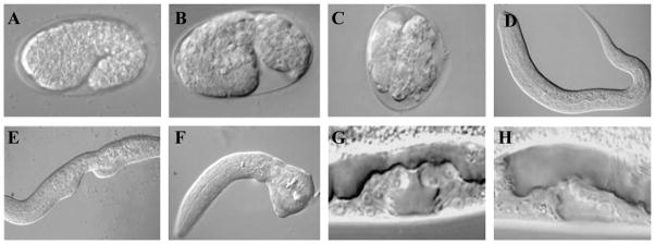
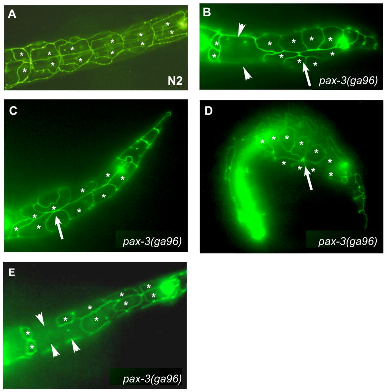
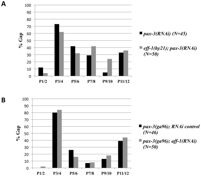
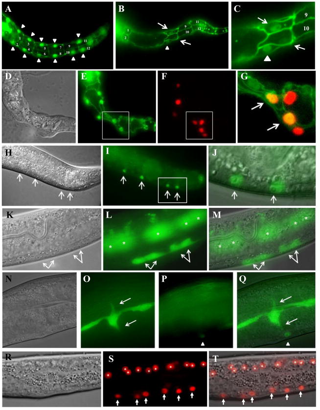
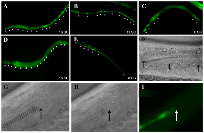
The development of the single cell layer skin or hypodermis of Caenorhabditis elegans is an excellent model for understanding cell fate specification and differentiation. Early in C. elegans embryogenesis, six rows of hypodermal cells adopt dorsal, lateral or ventral fates that go on to display distinct behaviors during larval life. Several transcription factors are known that function in specifying these major hypodermal cell fates, but our knowledge of the specification of these cell types is sparse, particularly in the case of the ventral hypodermal cells, which become Vulval Precursor Cells and form the vulval opening in response to extracellular signals. Previously, the gene pvl-4 was identified in a screen for mutants with defects in vulval development. We found by whole genome sequencing that pvl-4 is the Paired-box gene pax-3, which encodes the sole PAX-3 transcription factor homolog in C. elegans. pax-3 mutants show embryonic and larval lethality, and body morphology abnormalities indicative of hypodermal cell defects. We report that pax-3 is expressed in ventral P cells and their descendants during embryogenesis and early larval stages, and that in pax-3 reduction-of-function animals the ventral P cells undergo a cell fate transformation and express several markers of the lateral seam cell fate. Furthermore, forced expression of pax-3 in the lateral hypodermal cells causes them to lose expression of seam cell markers. We propose that pax-3 functions in the ventral hypodermal cells to prevent these cells from adopting the lateral seam cell fate. pax-3 represents the first gene required for specification solely of the ventral hypodermal fate in C. elegans providing insights into cell type diversification.

Keywords: C. elegans; Differentiation; Epidermis; Fate specification; Gene expression; PAX.


Conflict of interest statement

Conflict of Interest

The authors have no actual or potential conflict of interest including financial, personal or other relationships with other people or organizations that could inappropriately influence, or be perceived to influence, the work reported here.

Figures

Figure 1
Figure 1
Nomarski photomicrographs of wild-type and pvl-4(ga96) animals at different stages of development. (A) Wild-type 1.5 fold stage embryo. (B, C) pvl-4(ga96) embryos with disorganized morphology. (D) Wild-type early L1 stage larvae. (E) pvl-4(ga96) L1 larvae with Vab body morphology phenotype. (F) pvl-4(ga96) L1 larvae lacking posterior structures. (G) Wild-type L4 stage larvae with properly induced vulval structure (three Pn.p cells adopted induced fates). (H) pvl-4(ga96) L4 animal showing Underinduced vulval phenotype.
Figure 2
Figure 2. Transgenic rescue of pvl-4(ga96) phenotypes
pvl-4(ga96) animals were microinjected with a combination of pax-3p::pax-3::gfp (rescue) and ajm-1::gfp (marker) DNA. The extrachromosomal nature of the pax-3p::pax-3::gfp; ajm-1::gfp array allowed us to compare animals containing the array (marked by ajm-1::gfp expression in the pharynx and vulva) to animals not containing the array and subsequently score for rescue of pvl-4(ga96) phenotypes. (A–D) Nomarski images of L1 stage (A, B) and L4 stage (C, D) larvae. Animals containing the pax-3p::pax-3::gfp; ajm-1::gfp extrachromosomal array (green) were rescued for the Vab body morphology defect (B) and Underinduced vulval phenotype (D). Animals lacking the extrachromosomal array (non-green) showed these phenotypes (A, C). Note that the green color shows AJM-1::GFP at cell junctions in the pharynx (B) and developing vulva (D); PAX-3::GFP expression is too faint to see in these animals. Anterior is to the right and ventral towards the top in all images.
Figure 3
Figure 3. Embryonic and early larval expression of pax-3
(A–C) Nomarski (A), epifluorescent (B) and overlay (C) of embryo approximately 240 minutes post first cleavage expressing the pax-3p::mCherry reporter. (D) Schematic depicting the location of mCherry positive cells. Red cells indicate cells with mCherry expression in nuclei of labelled cells. Empty circles represent cells without pax-3 expression. Hypodermal cells are abbreviated with an h (e.g. h7 is hyp7). The P cells are labeled by brackets and numbered in pairs. The four symmetric pairs of cells in the posterior of the embryo are numbered as follows: (1) PVQ (2) PHB (3) T (4) hyp7. Red cells with a question mark were not identified. Note that pax-3p::mCherry reporter expression turns on in P cell pairs P1/2 and P9/10 just prior to the comma stage of embryogenesis. (E–H) Late comma stage embryo expressing the pax-3p::mCherry reporter and hypodermal cell junctional marker, ajm-1::gfp. AB-derived cells are labelled in H. (I) Ventral view of wild-type early L1-stage animal expressing ajm-1::gfp to mark hypdermal junctional boundaries. P cells are labeled 1–12 and are located between two rows of lateral seam cells (labelled V2-V6). (J) Early L1-stage animal expressing both ajm-1::gfp and pax-3p::mCherry reporters. At this stage the nuclei of the P cells have not migrated ventrally and are located near to the row of seam cells. (K) Late L1-stage animal with ajm-1::gfp, scm::gfp (marker of seam cell nuclei) and pax-3p::mCherry reporters. The a and p indicate anterior and posterior daughters of the indicated P cells, respectively. J and K are slightly ventro-lateral views, tilted left. The cells without mCherry expression in (J) and (K) are cells that do not express pax-3 reporters at this time (P3, P4, P11, P12) (L) Nomarski and (M) epifluorescence view of L4 stage vulva of animal carrying pax-3p::pax-3::gfp reporter, showing expression in VulF cells. Anterior is to the left.
Figure 4
Figure 4. pax-3 reduction-of-function causes P cell phenotypes
Epifluorescent images of L1-stage animals expressing the ajm-1::gfp hypodermal junctional marker. P cells are indicated with asterisks. (A) Wild-type L1 stage animal with six P cells pairs at the ventral midline. The seam cells are seen adjacent to P cells in a more dorsolateral position. (B – E) pax-3(ga96) L1-stage animals with P cells displaying either the Gap phenotype (loss of ajm-1::GFP expression between P cells: arrowheads) or the Disorganized phenotype (P cells making ectopic, inappropriate contacts with other P cells: arrows). All images are ventral views with anterior to the left.
Figure 5
Figure 5. pax-3 reduction-of-function causes P cell phenotypes
Prior to P cell pair separation and rotation it is not possible to distinguish individual P cells within pairs and the 12 P cells are grouped into six pairs (P1/2, P3/4, P5/6, P7/8, P9/10, and P11/12). All animals scored for the Gap or Dis phenotype (A, B) contained the ajm-1::gfp transgene. (A) Percentage of P cell pairs with Gap phenotype. (B) Percentage of P cell pairs with at least one cell displaying the Dis phenotype. (C) Percentage of P cell pairs in which at least one cell showed ectopic expression of scm::GFP in the P cells.
Figure 6
Figure 6. Reduction-of-function of the fusogen genes eff-1 and aff-1 function does not suppress the Gap P cell phenotype in pax-3 reduction-of-function animals
In A and B, L1-stage animals expressing ajm-1::gfp were scored for the percent occurrence of P cell pairs with cellular junction surrounding all four sides of P cells. (A) The Gap phenotype caused by pax-3 RNAi is not suppressed in an eff-1(hy21) mutant background. (B) aff-1 reduction-of-function by RNAi does not suppress pax-3(ga96) Gap P cell phenotype.
Figure 7
Figure 7. pax-3 reduction of function causes ectopic seam cell marker expression in P and Pn.p cells and misoriented seam cell processes
(A–C) Wild-type (A) and pax-3 RNAi treated animals (B, C) expressing ajm-1::gfp and scm::gfp. (C) is close up of image in (B). Arrowhead point to an scm::gfp seam cell; arrows point to P cells ectopically expressing scm::gfp. (D–G) pax-3(RNAi) on pax-3p::mCherry; ajm-1::gfp; scm::gfp early L1-stage animal. (D) Nomarski image, (E) ajm-1::gfp (junctional) and scm::gfp (nuclear) expression, (F) pax-3p::mCherry (nuclear) expression, (G) merge of boxed region in (E) and (F) showing coexpression of scm::gfp and pax-3p::mCherry in the same P cell nuclei (arrows). (H–J) Posterior end of an L3-stage pax-3(RNAi) animal with ectopic scm::gfp expression in Pn.p cells along the ventral surface. (H) Nomarski image, (I) fluorescent image, (J) merge of boxed region in (I). (K–M) L4-stage pax-3(RNAi) animal with ectopic grd-10p::gfp expression in P3.p and P4.p descendants (arrows). The cells marked by asterisks are the seam cells out of focus. (K) Nomarski image, (L) fluorescent image, (M) merge of (K) and (L). (N–Q) L4-stage pax-3(RNAi) animal with seam cells extending processes (arrows) towards Pn.p nuclei misexpressing grd-10::gfp (arrowhead). (N) Nomarski image, (O, P) fluorescent image in two different focal planes, (Q) merge of (N) and (O). (R–T) Posterior of an L2-stage pax-3(RNAi) animal with ectopic egl-18p::mCherry reporter expression in Pn.p cells (arrows). Asterisks mark seam cells after division. (R) Nomarski image, (S) fluorescent image, (T) merge of (R) and (S). Anterior is to the left and dorsal to the top in all images.
Figure 8
Figure 8. Ectopic pax-3 expression causes the loss of a seam cell marker and alae formation in seam cells
The grd-10p::gfp transgene shows leaky cytoplasmic expression making it possible to view both the nuclei and cell boundaries of the seam cells. (A) Non-heat-shocked hsp::pax-3; grd-10p::gfp animal with 16 seam cell nuclei expressing grd-10p::gfp. (B, C) hsp::pax-3; grd-10p::gfp animals given a single heat shock at the L2/L3 molt (B) and L3/L4 molt (C) showing 11 and 6 grd-10p::gfp expressing nuclei, respectively. (D) grd-10::gfp animal showing 16 grd-10::gfp expressing nuclei (E) grd-10p::pax-3; grd-10::gfp animals showing 8 grd-10::gfp expressing nuclei. (F) Nomarski image of wild-type animal showing continuous stretch of alae (arrows) (G–I) Nomarski (G, H) and epifluorescent (I) images of the same animal showing the loss of alae formation where grd-10p::gfp expression is absent (arrow). The four lines in G mark the alae seen in H. All images are of mid-L4-stage worms and anterior is to the left and dorsal towards the top. Asterisks mark individual seam cell nuclei. SC – seam cells nuclei with gfp expression.

References

    1. Andachi Y. Caenorhabditis elegans T-box genes tbx-9 and tbx-8 are required for formation of hypodermis and body-wall muscle in embryogenesis. Genes Cells. 2004;9:331–344. - PubMed
    1. Aspöck G, Kagoshima H, Niklaus G, Bürglin TR. Caenorhabditis elegans has scores of hedgehog-related genes: sequence and expression analysis. Genome Res. 1999;9:909–923. - PubMed
    1. Blake JA, Thomas M, Thompson JA, White R, Ziman M. Perplexing Pax: from puzzle to paradigm. Dev Dyn. 2008;237:2791–2803. - PubMed
    1. Blake JA, Ziman MR. Pax genes: regulators of lineage specification and progenitor cell maintenance. Development. 2014;141:737–751. - PubMed
    1. Brenner S. The genetics of Caenorhabditis elegans. Genetics. 1974;77:71–94. - PMC - PubMed

Publication types

MeSH terms

Substances

LinkOut - more resources