. 2016 Apr 22;60(5):2981-92.
doi: 10.1128/AAC.02932-15. Print 2016 May.

Enhancement of the Replication of Hepatitis C Virus Replicons of Genotypes 1 to 4 by Manipulation of CpG and UpA Dinucleotide Frequencies and Use of Cell Lines Expressing SECL14L2 for Antiviral Resistance Testing

Affiliations

Enhancement of the Replication of Hepatitis C Virus Replicons of Genotypes 1 to 4 by Manipulation of CpG and UpA Dinucleotide Frequencies and Use of Cell Lines Expressing SECL14L2 for Antiviral Resistance Testing

Jeroen Witteveldt et al. Antimicrob Agents Chemother. .

Abstract

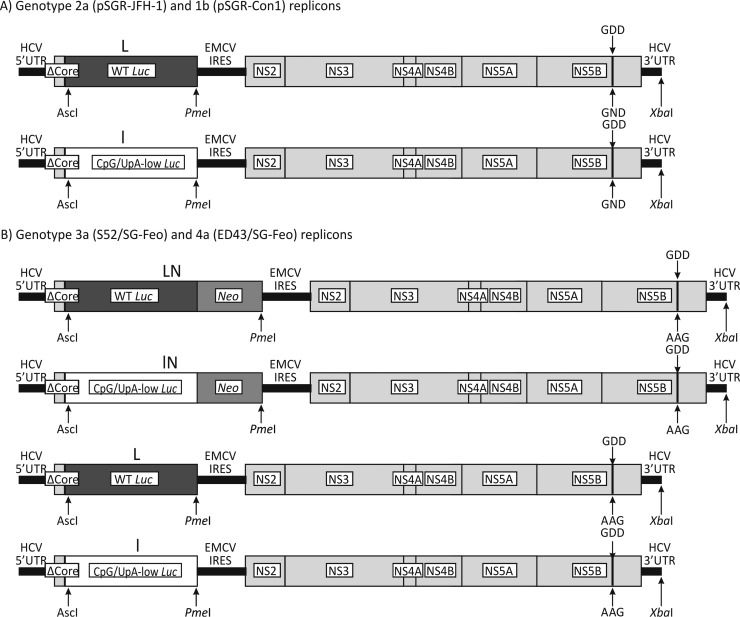
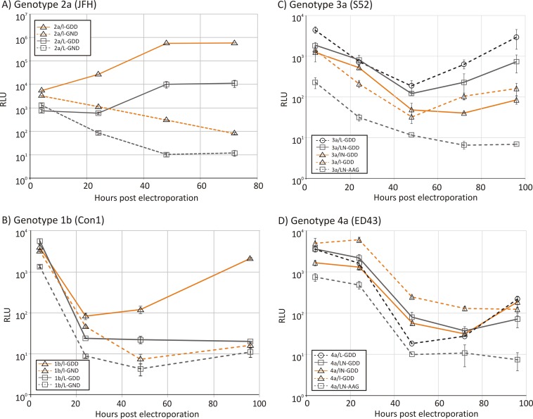
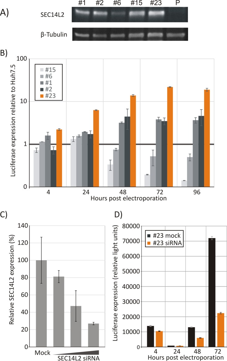
Treatment for hepatitis C virus (HCV) has improved greatly through the use of direct-acting antivirals (DAAs). However, their effectiveness and potential for drug resistance development in non-genotype 1 variants of HCV remain relatively unexplored, as in vitro assays to assess drug susceptibility are poorly developed and unsuited for a transient-transfection format. In the current study, we have evaluated the effects of dinucleotide frequency changes in the replicon and the use of a SEC14L2-expressing cell line on the replication of HCVs of different genotypes and evaluated the resulting assay formats for measurements of susceptibility to the DAA sofosbuvir. Removal of CpG and UpA dinucleotides from the luciferase gene used in HCV replicons of genotype 1b (Con1) and genotype 2a (JFH-1) achieved between 10- and 100-fold enhancement of replication over that of the wild type posttransfection. Removal of CpG and UpA dinucleotides in the neomycin gene or deletion of the whole gene in replicons of genotype 3a (S52) and genotype 4a (ED43) enhanced replication, but phenotypic effects on altering luciferase gene composition were minimal. A further 10-fold replication enhancement of replicons from all four genotypes was achieved by using a transgenic Huh7.5 cell line expressing SECL14L2, whose expression showed a dose-dependent effect on HCV replication that was reversible by small interfering RNA (siRNA) knockdown of gene expression. By combining these strategies, the 100- to 1,000-fold enhancement of replication allowed the susceptibility of all four genotypes to the RNA polymerase inhibitor sofosbuvir to be robustly determined in a transient-transfection assay format. These methods of replication enhancement provide new tools for monitoring the susceptibility and resistance of a wide range of HCV genotypes to DAAs.


Figures

FIG 1
FIG 1
Structures of subgenomic replicons and mutants. Shown are diagrammatic representations of the replicons used in this study, with changes to the luciferase gene (L and l), deletion of the neomycin gene (N), and replacement of the GDD motif in NS5B with GND (or AAG in genotypes 3 and 4) to make it replication defective labeled.
FIG 2
FIG 2
Replication of unmodified replicons and mutants with low-CpG/UpA luciferase. Shown are data for the replication of genotype 2a (A), 1b (B), 3a (C), and 4a (D) replicons after electroporation into Huh7.5 cells. Luciferase activity was measured at four time points (x axis) and plotted as absolute values (y axis). Error bars represent standard deviations. RLU, relative light units.
FIG 3
FIG 3
Expression of SEC14L2 in different cell lines and effect of HCV replication. (A) Quantification of SEC14L2 expression by Western blotting of cell lysates from different cell lines. Samples were normalized to the total amount of protein loaded; a control immunoblot using β-tubulin is shown at the bottom. P, parental Huh7.5 cell line. (B) Luciferase expression in different cell lines at different time points after electroporation of 1b/l-GDD. The values are normalized to luciferase expression of the 1b/l-GDD replicon in the parental Huh7.5 cell line at each time point. (C) Effect of transfection of SEC14L2-specific siRNA in cell line 23 on SEC14L2 expression levels as determined by Western blotting. (D) Replication of 1b/l-GDD in siRNA-treated and irrelevant siRNA-treated 23 cells. Error bars depict standard deviations.
FIG 4
FIG 4
Comparison of replication of replicons from genotypes 1 to 4 in the Huh7.5 and 23 cell lines. Shown are data for the replication of replicons from genotypes 1 to 4 generated in this study in the Huh7.5 and 23 cell lines; the right panels show luciferase expression from the replication-incompetent control. Error bars depict standard deviations.
FIG 5
FIG 5
In vitro translation assay of wild-type and low-CpG/UpA luciferase replicons. Shown are measurements of luciferase activity of translation products of WT and CpG/UpA-low HCV replicons of genotypes 1 to 4 after a 30-min incubation. Bars show the means of data from two replicates; error bars show standard deviations.
FIG 6
FIG 6
Coelectroporation of CpG/UpA-low and WT luciferase-containing replicons. Shown are luciferase expression levels in Huh7.5 cells (A) and 23 cells (B) after electroporation of CpG/UpA-low, WT, or both replicons; the y-axis scale is normalized to luciferase expression by 1b/l-GDD (100%). Error bars depict standard deviations.
FIG 7
FIG 7
Testing of susceptibility of original and modified replicons to sofosbuvir (Sofos.). Shown are data for the inhibition of replication of CpG-low and WT luciferase replicons 1b/l-GDD and 1b/L-GDD (A), 3a/LN-GDD and 3a/L-GDD (B), and 4a/LN-GDD and 4a/l-GDD (C) with differing concentrations of sofosbuvir. The experiment was performed with the Huh7.5 and 23 cell lines. Error bars depict standard deviations.

Similar articles

Cited by

References

    1. Choo QL, Kuo G, Weiner AJ, Overby LR, Bradley DW, Houghton M. 1989. Isolation of a cDNA clone derived from a blood-borne non-A, non-B viral hepatitis genome. Science 244:359–362. doi:10.1126/science.2523562. - DOI - PubMed
    1. Alter MJ. 1991. Hepatitis C: a sleeping giant? Am J Med 91:112S–115S. doi:10.1016/0002-9343(91)90354-Z. - DOI - PubMed
    1. Bell J, Batey RG, Farrell GC, Crewe EB, Cunningham AL, Byth K. 1990. Hepatitis C virus in intravenous drug users. Med J Aust 153:274–276. - PubMed
    1. Hanafiah KM, Groeger J, Flaxman AD, Wiersma ST. 2013. Global epidemiology of hepatitis C virus infection: new estimates of age-specific antibody to HCV seroprevalence. Hepatology 57:1333–1342. doi:10.1002/hep.26141. - DOI - PubMed
    1. Messina JP, Humphreys I, Flaxman A, Brown A, Cooke GS, Pybus OG, Barnes E. 2015. Global distribution and prevalence of hepatitis C virus genotypes. Hepatology 61:77–87. doi:10.1002/hep.27259. - DOI - PMC - PubMed

Publication types