Review
. 2016 May 8;428(9 Pt A):1659-80.
doi: 10.1016/j.jmb.2016.02.027. Epub 2016 Mar 4.

Mechanisms of Selective Autophagy in Normal Physiology and Cancer

Affiliations
Review

Mechanisms of Selective Autophagy in Normal Physiology and Cancer

Joseph D Mancias et al. J Mol Biol. .

Abstract

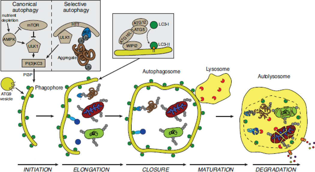
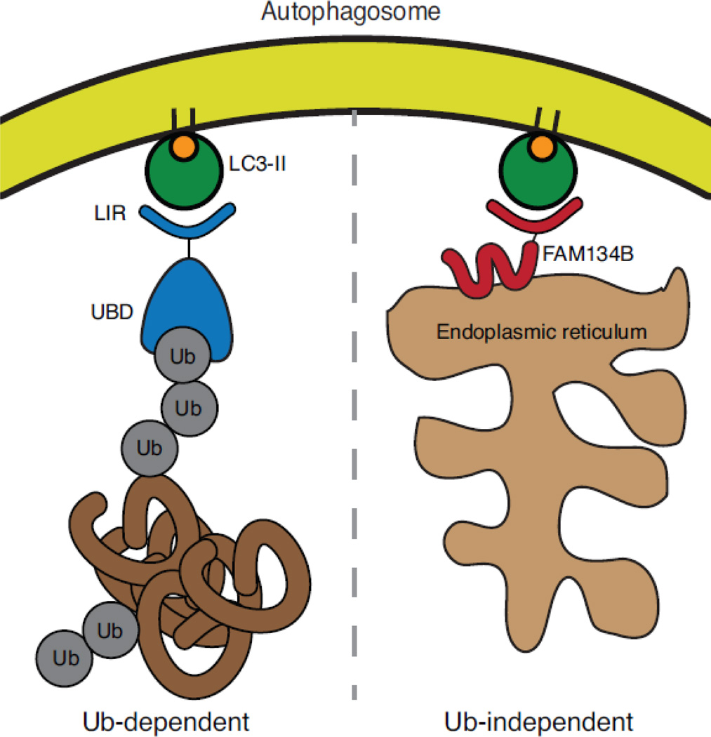
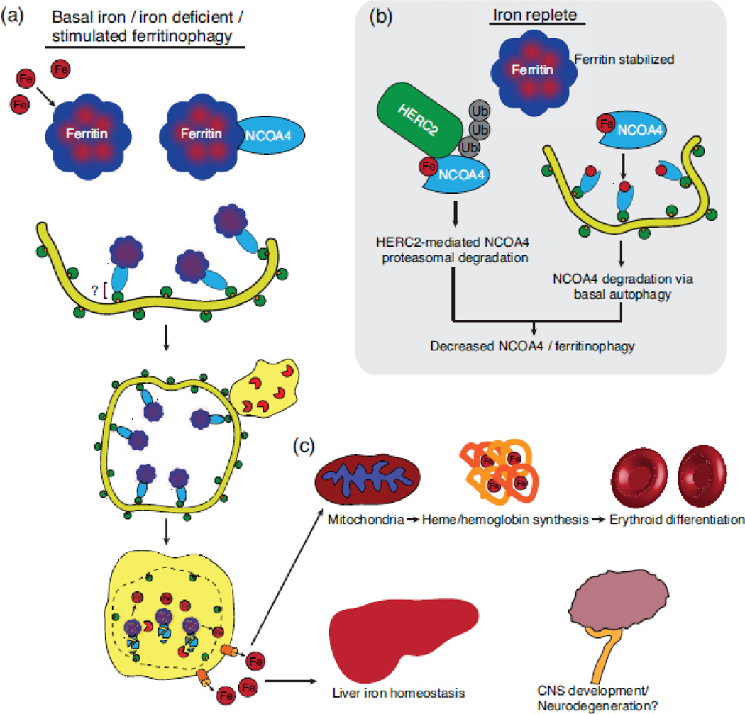
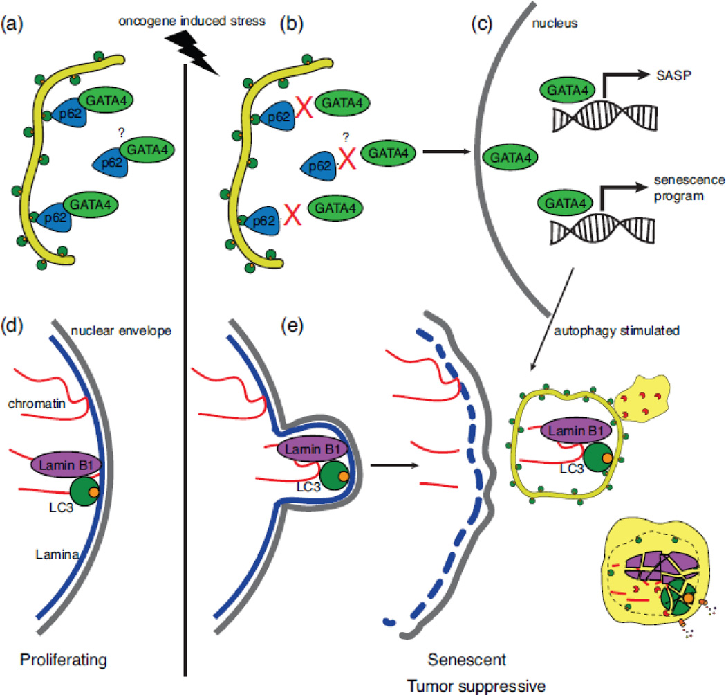
Selective autophagy is critical for regulating cellular homeostasis by mediating lysosomal turnover of a wide variety of substrates including proteins, aggregates, organelles, and pathogens via a growing class of molecules termed selective autophagy receptors. The molecular mechanisms of selective autophagy receptor action and regulation are complex. Selective autophagy receptors link their bound cargo to the autophagosomal membrane by interacting with lipidated ATG8 proteins (LC3/GABARAP) that are intimately associated with the autophagosome membrane. The cargo signals that selective autophagy receptors recognize are diverse but their recognition can be broadly grouped into two classes, ubiquitin-dependent cargo recognition versus ubiquitin-independent. The roles of post-translational modification of selective autophagy receptors in regulating these pathways in response to stimuli are an active area of research. Here we will review recent advances in the identification of selective autophagy receptors and their regulatory mechanisms. Given its importance in maintaining cellular homeostasis, disruption of autophagy can lead to disease including neurodegeneration and cancer. The role of autophagy in cancer is complex as autophagy can mediate promotion or inhibition of tumorigenesis. Here we will also review the importance of autophagy in cancer with a specific focus on the role of selective autophagy receptors.

Keywords: cancer; ferritinophagy; macroautophagy (autophagy); mitophagy; selective autophagy.


Figures

Figure 1
Figure 1
The process and regulation of selective autophagy. The stages of autophagy (initiation, elongation, closure, maturation, and degradation) are shown. Cargo is sequestered in selective and bulk degradative manners via a double-membrane phagophore that fuses on itself to form the autophagosome. This subsequently fuses to the lysosome (autolysosome), where the cargo is degraded by lysosomal enzymes and degradation products are recycled to the cytosol by lysosomal transporters[198]. mTOR is a key regulator of bulk autophagy in response to changes in nutrient availability. During nutrient-replete conditions, mTOR is activated and autophagy is inhibited through repression of ULK1/2 (the mammalian homologs of ATG1). Upon nutrient depletion, the ULK1/2 complex is activated and can promote autophagy initiation. ULK1/2 is also activated at low energy states (an increased AMP/ATP ratio) by phosphorylation via AMPK as well as repression of mTORC1 activity. Activation of the selective autophagy pathway is via multiple specific stimuli and how and whether these stimuli engage the ULK complex in all circumstances is less well understood. Huntingtin (mammalian homology of yeast ATG11) was recently identified as a scaffold protein that can activate the ULK complex and bring a selective autophagy receptor into close proximity with activated ULK complex thereby linking activation of autophagosome formation with cargo destined for degradation. Also critical to autophagy initiation is the production of phosphatidylinositol-3-phosphate (PI3P) by the class III PI3K complex, here labeled ‘PI(3)KC3’ composed of Vps34, ATG14, ATG6/Beclin1 and p150 (Vps15). ATG9 containing vesicles contribute membrane to the growing autophagosome and likely participate in recruitment of other essential autophagy machinery (not shown)[29]. WIPI2 binds ATG16L1 to localize the ATG5–ATG12-ATG16L1 complex to autophagosomal membranes. This complex acts downstream of the ULK and PI(3)KC3 complexes in an E3-like manner to conjugate phosphatidylethanolamine (PE) to LC3-I to produce lipidated LC3-II that then associates with autophagosomal membranes and has roles in autophagosome membrane elongation. LC3-II is present on the outer and inner surfaces of the autophagosome (depicted as a green circle with orange PE moiety) and acts as the physical link between selective autophagy receptors and the autophagosome membrane. Selective autophagic cargos depicted here include ubiquitylated mitochondria, ubiquitylated bacteria (light green rounded rectangle), ubiquitylated protein aggregates recognized by a selective autophagy receptor (brown tangle), and nonubiquitylated cargo, such as ferritin (blue circle recognized by light blue oval depicting NCOA4).
Figure 2
Figure 2
Ubiquitin-dependent and ubiquitin–independent selective autophagy. On the left, a prototypical selective autophagy receptor with an ubiquitin-binding domain (UBD) recognizes an ubiquitylated substrate (protein aggregate) and physically links the aggregate to the autophagosome via a LIR motif that binds to lipidated and autophagosome membrane associated ATG8 (LC3-II). On the right, FAM134B-mediated endoplasmic reticulum-phagy (ER-phagy), a ubiquitin-independent selective autophagy pathway is depicted. Here, FAM134B is an ER-membrane protein with a LIR motif that is recognized by autophagosome associated LC3-II in order to deliver predominantly cisternal ER to autophagosomes for degradation.
Figure 3
Figure 3
NCOA4-mediated ferritinophagy pathway. (a) Iron (Fe) is sequestered in 24-mer ferritin complexes containing ferritin heavy and light chains. NCOA4 recognizes ferritin and delivers it to an incipient autophagosome. The molecular nature of the NCOA4-autophagosome interaction is unclear but likely involves interaction with LC3-II potentially via a non-canonical LIR motif. Degradation of ferritin in an autolysosome releases iron, which is exported to the cytosol. (b) NCOA4 levels and thereby ferritinophagy are regulated by iron levels in the cell. Under high iron/iron replete conditions, NCOA4 is recognized by HERC2, an E3 ubiquitin ligase, in an iron-dependent manner and targeted for proteasomal degradation. In tandem, NCOA4 is also targeted for autophagic degradation. A lower level of NCOA4 therefore decreases flux through the ferritinophagy pathway. (c) Liberated iron can be used in many iron-dependent processes including heme synthesis, which is required for hemoglobin synthesis during erythroid differentiation. Furthermore, NCOA4-mediated ferritinophagy is important for other physiological processes such as maintenance of liver iron homeostasis. Further research will be required to understand the role of NCOA4 in CNS development and pathophysiology of iron-associated neurodegenerative disorders.
Figure 4
Figure 4
Selective autophagy in cancer. (a) Cellular senescence is a barrier to tumorigenesis and selective autophagy explains the conflicting reports about the role of autophagy in senescence. Normal proliferating cells basally degrade the pro-senescence transcription factor GATA4 via p62 selective autophagy. The molecular details of the GATA4-p62 interaction and its regulation are currently unclear. (b) Upon oncogene-induced stress, the GATA4-p62 interaction is abrogated by an unknown mechanism thereby stabilizing GATA4. (c) GATA4 translocates to the nucleus where it activates a senescence transcriptional program as well as a senescence associated secretory phenotype (SASP) transcriptional program. General autophagy is also stimulated at this time and is required to drive senescence. This pathway clarifies the apparent paradox in the literature that autophagy can suppress senescence (basal selective autophagy of GATA4) and promote senescence (autophagy is required to support senescence phenotype). (d) Basally, Lamin B1 binds nuclear LC3 and chromatin but is not targeted for autophagic degradation. (e) Upon oncogene-induced stress, Lamin B1 is autophagocytosed with bound chromatin to mediate nuclear lamina selective autophagy contributing to a senescence phenotype and thereby tumor suppression.

References

    1. Yang Z, Klionsky DJ. Eaten alive: a history of macroautophagy. Nature Cell Biology. 2010;12:814–822. - PMC - PubMed
    1. Kimmelman AC. The dynamic nature of autophagy in cancer. Genes & Development. 2011;25:1999–2010. - PMC - PubMed
    1. Mizushima N, Yoshimori T, Ohsumi Y. The Role of Atg Proteins in Autophagosome Formation. Annu. Rev. Cell Dev. Biol. 2011;27:107–132. - PubMed
    1. Noda NN, Inagaki F. Mechanisms of Autophagy. Annu. Rev. Biophys. 2015;44:101–122. - PubMed
    1. Svenning S, Johansen T. Selective autophagy. Essays Biochem. 2013;55:79–92. - PubMed

Publication types

LinkOut - more resources