. 2016 Mar 7;36(5):487-97.
doi: 10.1016/j.devcel.2016.02.008.

HP1-Assisted Aurora B Kinase Activity Prevents Chromosome Segregation Errors

Affiliations

HP1-Assisted Aurora B Kinase Activity Prevents Chromosome Segregation Errors

Yusuke Abe et al. Dev Cell. .

Abstract

Incorrect attachment of kinetochore microtubules is the leading cause of chromosome missegregation in cancers. The highly conserved chromosomal passenger complex (CPC), containing mitotic kinase Aurora B as a catalytic subunit, ensures faithful chromosome segregation through destabilizing incorrect microtubule attachments and promoting biorientation of chromosomes on the mitotic spindle. It is unknown whether CPC dysfunction affects chromosome segregation fidelity in cancers and, if so, how. Here, we show that heterochromatin protein 1 (HP1) is an essential CPC component required for full Aurora B activity. HP1 binding to the CPC becomes particularly important when Aurora B phosphorylates kinetochore targets to eliminate erroneous microtubule attachments. Remarkably, a reduced proportion of HP1 bound to CPC is widespread in cancers, which causes an impairment in Aurora B activity. These results indicate that HP1 is an essential modulator for CPC function and identify a molecular basis for chromosome segregation errors in cancer cells.


Figures

Figure 1
Figure 1. Characterization of HP1 association with the CPC in mitosis
(A) HP1 proteins localize to centromeres in mitosis. Immunofluorescence microscopy of mitotic chromosomes from HeLa cells stained for HP1α, HP1β, HP1γ or Aurora B. Scale bar indicates 10 μm. (B) All three types of HP1 coimmunoprecipitate with the CPC. The CPC from mitotic HeLa cell extracts, with or without depletion of HP1, was immunoprecipitated using INCENP antibodies. Note that HP1 proteins bind to the CPC in a compensatory manner. (C) The amount of HP1 proteins stably bound to CPC. Quantities of the indicated proteins in immunoprecipitates of CPC from mitotic HeLa cell extracts were estimated by comparing to their recombinant proteins of known concentrations (Figures S1C–H). The proportion of HP1-bound CPC is calculated assuming that “dimerized” HP1 proteins associate with the CPC. (D) The CPC was immunoprecipitated from mitotic RPE1 and HeLa cell extracts using INCENP antibodies. Comparable amounts of INCENP complexes from both cell lines were assessed for co-immunoprecipitated HP1 proteins. The HP1:INCENP ratio in protein complexes was compared in HeLa and RPE1 cells (The ratio in RPE1 cells is set to 1.0). (E) Mitotic chromosome spreads from RPE1 and HeLa cells were costained with antibodies to HP1α, HP1β, or HP1γ and INCENP. Relative fluorescence intensities of HP1α, HP1β, or HP1γ, normalized to INCENP, are shown in the histogram. n > 140 centromeres from 4 cells were analyzed. Error bars indicate SEM. Scale bar indicates 10 μm.
Figure 2
Figure 2. Proportion of HP1-bound CPC is reduced in cancer cells
(A) The CPC was immunoprecipitated with INCENP antibodies from mitotic cell extracts of various types of cell lines, and immunoblotted with the indicated antibodies. The amount of co-immunoprecipitated HP1 proteins in RPE1 cells is set to 1.0 in the histogram. (B) Proportion of HP1-bound CPC is analyzed in cancer cell lines characterized with modest chromosome missegregation rates (HCT 116 and DLD-1) as in (A).
Figure 3
Figure 3. HP1 is required to attain mitotic activity of Aurora B in full
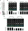
(A) Depletion of HP1 from the CPC. Three HP1 subtypes (HP1αβγ) were simultaneously depleted from HeLa cells using RNA interference (RNAi)-mediated knockdown, and the CPC was immunoprecipitated with antibodies to INCENP. (B and C) Immunofluorescence microscopy of Aurora B-mediated phospho-proteins. Following HP1 triple knockdown (HP1αβγ) or treatment with either a low dose (0.4 μM) or high-dose (2.0 μM) of Aurora B inhibitor ZM447439 (ZM), HeLa cells were fixed and stained with antibodies to H3 phospho-Ser10, pS10 (B), to Hec1 phospho-Ser44, pS44 and to Hec1 that do not distinguish phosphorylation status (Hec1) Relative fluorescence intensities of Hec1-pS44 are normalized to Hec1, and are summarized in the histogram. n >150 kinetochores from 10 cells were analyzed for each condition; error bars indicate SEM (C). Representative cells in prometaphase are shown. Scale bar indicates 5 μm. (D) HP1α domain structure, highlighting the conserved mitotic phosphorylation site Ser92 in the hinge domain (left panel). A single alanine mutation at this site, S92A, significantly inhibits its Aurora B-mediated phosphorylation in vitro (right panel). (E) Antibodies to HP1α phosphor-Ser92 (HP1α-pS92) preferentially immunoprecipitate HP1-bound CPC from mitotic HeLa cell extracts. A serial dilution of the sample indicates that far more HP1 proteins were immunoprecipitated with the CPC with HP1α-pS92 antibodies (left panel). Both CPC populations were assayed for kinase activity (right panel). Note that most part of the kinase activity was sensitive to Aurora B inhibitor (+ ZM).
Figure 4
Figure 4. HP1 assists the activity of Aurora B by increasing its affinity to substrates
(A) Schematic diagram of INCENP mutants (left). The sequence targeted by RNAi, in which RNAi-resistant substitutions were introduced into cDNA, is highlighted in blue. Immunoprecipitation with myc antibodies from mitotic extract prepared from WT or ΔHP INCENP expressing HeLa cells (right). Note that the ΔHP mutant can assemble the complex but largely lacks the ability to bind to all three HP1 subtypes. (B) Mitotic HeLa cell extracts expressing myc-tagged WT, ΔHP or 3A INCENP (left panel) were used to immunoprecipitate the CPC using antibodies to myc (middle panel) and assayed for Aurora B kinase activity toward GST-Hec1 (1-80) (right panel). The graph shows mean kinase activity of six independent experiments (error bars indicate SD). *P < 0.01, two-tailed Student’s t-test. Note that the ΔHP mutant seemed otherwise functional because the generation of multinuclear cells after INCENP depletion (Hauf et al., 2003; Ditchfield et al., 2003) was rescued by ΔHP INCENP expression (Figure S4F). An in vitro kinase assay with recombinant ΔHP INCENP-Aurora B verifies that Aurora B activity is not compromised (Figure S4L). (C) Kinase reaction using recombinant proteins. Co-purified INCENP (full length)−Aurora B was used as an enzyme source, in the presence or absence of preincubation with HP1α, and phosphate incorporation on GST-Hec1(1-80) was measured after indicated times of reaction. (D) INCENP−Aurora B kinase assay was performed following preincubation with all three subtypes of HP1 proteins. Note that HP1α and HP1γ, but not HP1β, are phosphorylated. Enhancement of kinase activity was similarly seen toward histone H3 (Figure S4I). (E) INCENP−Aurora B preincubated with indicated HP1α proteins (upper panel) were assayed for their kinase activity as in (C) (lower panel). Enhancement of kinase activity was also seen toward histone H3 (Figure S4J). (F) Two CPC populations, i.e., with or without enrichment of HP1-bound, were analyzed for Aurora B and INCENP phosphorylation status by Phos-tag gel electrophoresis and phosphospecific antibodies (INCENP-pTSS), respectively. (G) Kinetic profile of Aurora B kinase activity in the presence or absence of HP1α with different concentrations of substrate. Phosphate incorporation to GST-Hec1(1-80) was measured and plotted. Experiments were performed in triplicate and data represent mean values ± SEM. Note that the kcat value for the GST-Hec1(1-80) substrates in the presence of HP1α is significantly higher than that in the absence of HP1α. (H) Binding assay between the kinase and substrate. Co-purified recombinant INCENP full-length and GST-Aurora B were incubated with His-tagged Hec1(1-80) in the presence or absence of indicated HP1α proteins (input), and resulting precipitates with GST-tag were analyzed by immunoblotting (GST pulldown). Graph shows the relative amount of Hec1(1-80)-His in the precipitate from five independent experiments (bars indicate SD). *P < 0.01, two-tailed Student’s t-test.
Figure 5
Figure 5. The HP1-mediated allosteric regulation is essential for Aurora B-mediated phosphorylation of kinetochore substrates
(A) Fluorescence intensities of Hec1-pS44 in WT or ΔHP INCENP-expressing HeLa cells. Relative fluorescence intensities are summarized as in Figure 3C. n >150 kinetochores from 10 cells for each condition were analyzed; error bars indicate SEM. (B) The centromere localization of all three subtypes of HP1 proteins depends on their binding to INCENP. Chromosome spreads from mitotic INCENP WT or ΔHP expressing HeLa cells, with or without INCENP depletion, were stained with antibodies to HP1α, HP1β and HP1γ. Scale bar indicates 10 μm. (C) Immunofluorescence microscopy of Hec1-pS44 in HeLa cells expressing CENP-B-fused INCENP (CB-INCENP-EGFP), either WT or ΔHP mutant, with or without RNAi treatment to deplete endogenous INCENP. Fluorescence intensities of Hec1-pS44 are summarized in the histogram. n >150 kinetochores from more than 8 cells were analyzed; error bars indicate SEM; scale bar, 5 μm.
Figure 6
Figure 6. Sufficient levels of Aurora B activity in non-transformed cells depends largely on HP1-mediated allosteric regulation of the CPC
(A) INCENP-associated kinase activity in mitotic RPE1, HT-1080, and U2OS cells. Kinase activity is analyzed for equivalent amounts of INCENP immunoprecipitated from three cell lines. Graph shows mean kinase activity from six independent experiments (error bars indicate SD). *P < 0.01, two-tailed Student’s t-test. (B and C) Dsn1 phosphorylation levels are elevated in non-transformed cells than in cancer cells. Immunofluorescence microscopy of Dsn1-pS100 and Hec1 (B) or Dsn1 and Hec1 (C) in cancer (HeLa and U2OS) or non-transformed (RPE1 and TIG-3) cell lines. Relative fluorescence intensities of Dsn1-pS100 or Dsn1, both being normalized to Hec1, are shown in the histogram. n > 250 kinetochores from more than 10 cells for each condition were analyzed. Error bars indicate SEM; scale bar, 5 μm. (D) Immunofluorescence microscopy of Dsn1-pS100 in WT or ΔHP INCENP-expressing HeLa and RPE1 cells, in the presence or absence of endogenous INCENP. Relative fluorescence intensities are summarized as in (B). n > 300 kinetochores from 14 cells for each condition; error bars indicate SEM; scale bar 5μm.
Figure 7
Figure 7. HP1 binding to the CPC is required to prevent chromosome segregation errors in non-transformed cells
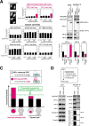
(A) Cells stably expressing WT or ΔHP INCENP were generated in non-transformed (RPE1 and TIG-3; Figures S6A, B) and cancer (HeLa, U2OS, HT-1080, A549, HCT 116 and DLD-1; Figures S6C–G) cell lines. Chromosome missegregation rates in anaphase were calculated with or without knockdown of endogenous INCENP from more than 200 cells in unperturbed mitoses or in cells that had been released from Monastrol treatment (Figure S7A). The histogram shows the mean ± SD of three independent experiments. Similar results were obtained in two other independent subclones of RPE1 cells expressing ΔHP INCENP (Figure S7B–D), and also in subclones of RPE1 (Figure S7E–G) and HeLa (Figure S7H; characterized in Figure 4B) expressing 3A INCENP. (B) CPCs were immunoprecipitated using antibodies to INCENP from mitotic extracts of parental HeLa cells and from cells engineered to conditionally overexpress GFP-tagged HP1α, and blotted for HP1 proteins. Graph shows the relative amount of HP1 referenced to INCENP in the precipitates. The amount of endogenous HP1 proteins in parental cells was set to 1.0. (C) A diagram depicting composition of the CPC in non-transformed and cancer cell lines. Non-transformed cells contain more HP1-bound CPC than cancer cells do, and thus the former has higher Aurora B kinase activity than the latter. The HP1-bound CPC is particularly important to phosphorylate kinetochore substrates and thus to destabilize incorrect microtubule attachments in non-transformed cells. In wide range of cancer cells, however, proportion of HP1-bound CPC and Aurora kinase B activity are reduced beyond the threshold required to prevent chromosome segregation errors. (D) Generation of transformed RPE1-67R cell lines (upper panel). Mitotic extracts from parental RPE1 cells and transformed RPE1-67R cells (input) were analyzed as in (B). Phospho-H3 Ser10 bands confirm the mitotic state. The amount of HP1 in INCENP complexes in RPE1 cells is set to 1.0 in the histogram.

Comment in

References

    1. Ainsztein AM, Kandels-Lewis SE, Mackay AM, Earnshaw WC. INCENP centromere and spindle targeting: identification of essential conserved motifs and involvement of heterochromatin protein HP1. J Cell Biol. 1998;143:1763–1774. - PMC - PubMed
    1. Bakhoum SF, Silkworth WT, Nardi IK, Nicholson JM, Compton DA, Cimini D. The mitotic origin of chromosomal instability. Curr Biol. 2014;24:R148–R149. - PMC - PubMed
    1. Bishop JD, Schumacher JM. Phosphorylation of the carboxyl terminus of inner centromere protein (INCENP) by the Aurora B Kinase stimulates Aurora B kinase activity. J Biol Chem. 2002;277:27577–27580. - PMC - PubMed
    1. Brasher SV, Smith BO, Fogh RH, Nietlispach D, Thiru A, Nielsen PR, Broadhurst RW, Ball LJ, Murzina NV, Laue ED. The structure of mouse HP1 suggests a unique mode of single peptide recognition by the shadow chromo domain dimer. EMBO J. 2000;19:1587–1597. - PMC - PubMed
    1. Carmena M, Wheelock M, Funabiki H, Earnshaw WC. The chromosomal passenger complex (CPC): from easy rider to the godfather of mitosis. Nat Rev Mol Cell Biol. 2012;13:789–803. - PMC - PubMed

Publication types

MeSH terms

Substances