. 2016 Mar 8;12(3):e1005498.
doi: 10.1371/journal.ppat.1005498. eCollection 2016 Mar.

Cyclin-Dependent Kinase CRK9, Required for Spliced Leader trans Splicing of Pre-mRNA in Trypanosomes, Functions in a Complex with a New L-Type Cyclin and a Kinetoplastid-Specific Protein

Affiliations

Cyclin-Dependent Kinase CRK9, Required for Spliced Leader trans Splicing of Pre-mRNA in Trypanosomes, Functions in a Complex with a New L-Type Cyclin and a Kinetoplastid-Specific Protein

Nitika Badjatia et al. PLoS Pathog. .

Abstract

In eukaryotes, cyclin-dependent kinases (CDKs) control the cell cycle and critical steps in gene expression. The lethal parasite Trypanosoma brucei, member of the phylogenetic order Kinetoplastida, possesses eleven CDKs which, due to high sequence divergence, were generically termed CDC2-related kinases (CRKs). While several CRKs have been implied in the cell cycle, CRK9 was the first trypanosome CDK shown to control the unusual mode of gene expression found in kinetoplastids. In these organisms, protein-coding genes are arranged in tandem arrays which are transcribed polycistronically. Individual mRNAs are processed from precursor RNA by spliced leader (SL) trans splicing and polyadenylation. CRK9 ablation was lethal in cultured trypanosomes, causing a block of trans splicing before the first transesterification step. Additionally, CRK9 silencing led to dephosphorylation of RNA polymerase II and to hypomethylation of the SL cap structure. Here, we tandem affinity-purified CRK9 and, among potential CRK9 substrates and modifying enzymes, discovered an unusual tripartite complex comprising CRK9, a new L-type cyclin (CYC12) and a protein, termed CRK9-associated protein (CRK9AP), that is only conserved among kinetoplastids. Silencing of either CYC12 or CRK9AP reproduced the effects of depleting CRK9, identifying these proteins as functional partners of CRK9 in vivo. While mammalian cyclin L binds to CDK11, the CRK9 complex deviates substantially from that of CDK11, requiring CRK9AP for efficient CRK9 complex formation and autophosphorylation in vitro. Interference with this unusual CDK rescued mice from lethal trypanosome infections, validating CRK9 as a potential chemotherapeutic target.


Conflict of interest statement

The authors have declared that no competing interests exist.

Figures

Fig 1
Fig 1. Tandem affinity purification of CRK9.
(A) Schematic depiction (not to scale) of the CRK9 locus in procyclic TbC9ee cells that exclusively express CRK9-PTP and no untagged CRK9. In these cells one wild-type CRK9 allele (open box) was knocked out by a hygromycin phosphotransferase gene (HYG-R, striped box). Integration of plasmid CRK9-PTP-NEO introduced the PTP sequence (black box) and the neomycin phosphotransferase (NEO-R) to the second allele. Smaller gray boxes indicate gene flanks for RNA processing signals and checkered boxes depict CRK9 sequences encoded in the plasmid. (B) Immunoblot of whole cell lysates of wild-type trypanosomes and of TbC9ee cells, detecting CRK9 and CRK9-PTP, respectively, with the newly generated anti-CRK9 polyclonal immune serum. On the same blot, phosphorylated (p) and unphosphorylated RPB1 was detected as a loading control with a polyclonal antibody, and CRK9-PTP specifically with the peroxidase anti-peroxidase reagent (PAP) that binds to the ProtA domains of the PTP tag. (C) Immunoblot monitoring of the CRK9-PTP purification detecting CRK9-PTP in crude extract (Inp) and the flowthrough of IgG affinity chromatography (FT-IgG), and CRK9-P in TEV protease eluate, the flowthrough of the anti-ProtC immunoaffinity chromatography (FT-ProtC) and the final eluate (Elu) with the monoclonal HCPC4 anti-ProtC antibody. The x-values indicate relative amounts analyzed. (D) Protein analysis of the CRK9-PTP purification. Proteins of crude extract, the TEV eluate and the final eluate were separated on 10–20% SDS polyacrylamide gel and first stained with SYPRO Ruby and, subsequently, with Coomassie blue (right panel). Marker sizes in kDa are indicated on the left.
Fig 2
Fig 2. CRK9 interacts and co-sediments with two unannotated proteins.
(A) CRK9-PTP tandem affinity-purified material was sedimented through a 10–40% linear sucrose gradient by ultracentrifugation and fractionated into 20 aliquots from top to bottom. Note that pelleted proteins were resuspended in fraction 20 (20+P). Proteins from each fraction were separated by SDS-PAGE and stained with SYPRO Ruby. Protein bands were excised and identified by LC/MS/MS. Arrows point to the CYC12 and CRK9AP bands which co-sediment with CRK9 in fractions 9/10. The 35 kDa band with a peak in fractions 10/11 was found to be the putative ribosomal protein L5 (Tb927.9.15110/15150). (B) Kinase assay with materials from indicated fractions suggest autophosphorylation of CRK9. (C) Reciprocal co-IP assays of extracts prepared from a cell line in which CRK9 was exclusively PTP-tagged and an HA tag sequence was inserted at the 3’end of one CYC12 allele. The precipitate (P) was loaded at a fourfold excess relative to extract (Inp) and supernatant (S). Detection of the RNA pol II transcription factor TFIIB served as a negative precipitation control.
Fig 3
Fig 3. Cyclin CYC12 is an L-type cyclin.
(A) Schematic drawing to scale of the human L1 and T. brucei CYC12 cyclins. The two cyclin folds (blue) are embedded in the CCL1 domain (green). The charged RS domain (red) was defined by a hydrophilicity Kyte & Doolittle blot score of < -2. Black lines indicate SR or RS dipeptides. Both cyclin domains of CYC12 are disrupted by insertions. (B) Phylogenetic tree, generated by the maximum likelihood algorithm and based on a multiple sequence alignment of the cyclin domains of human cyclins and of CYC12s from T. brucei (Tb), T. cruzi (Tc), L. major (Lm) and the bodonid Bodo saltans (Bs) (for accession numbers see S5 Fig). Cyclins involved in the cell cycle and in transcriptional control are indicated. Bootstrap values are indicated in percentages and were derived from 1000 replicates. The common branch of human cyclins L and kinetoplastid CYC12s is drawn in red.
Fig 4
Fig 4. CYC12 and CRK9AP are functional partners of CRK9.
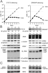
(A) Cumulative culture growth curves were obtained for CYC12 and CRK9AP silencing in the absence and presence of doxycycline (dox), the gene knockdown-inducing compound. For each knockdown a representative growth curve is shown. (B) Analysis of total RNA prepared from non-induced cells and cells in which CYC12 or CRK9AP were silenced for 1, 2 or 3 days. CYC12 or CRK9AP mRNA as well as α tubulin and RPB7 mRNA were analyzed by reverse transcription of oligo-dT and semi-quantitative PCR, whereas unspliced, pre-mRNA of α tubulin and RPB7 were analyzed by reverse transcription of random hexamers and by PCR using an oligonucleotide upstream of the SL addition site. rRNA was visualized by ethidium bromide staining after separation in an agarose gel. SL RNA, U2 snRNA and the Y structure intermediate were detected by primer extension assays using a SL RNA and a U2 snRNA-specific primer in the same reactions. (C) Anti-RPB1 immunoblot analysis of whole-cell lysates prepared from CRK9AP-silenced cells. Detection of the similar-sized RNA pol I subunit RPA1 served as a loading control.
Fig 5
Fig 5. CRK9AP depletion results in rapid co-loss of CRK9 and CYC12.
(A) Immunoblot of whole cell lysates derived from non-induced (n.i.) and CRK9AP-silenced PF trypanosomes. The arrow indicates the gene knockdown of CRK9AP. Detection of the class I transcription factor A subunit 6 (CITFA6) served as a loading control. (B) Corresponding semi-quantitative PCR analysis of cDNA that was obtained from the same cells by reverse transcription of total RNA using oligo-dT. Relative RNA amounts were determined by ethidium bromide–stained rRNA.
Fig 6
Fig 6. CRK9AP is essential for CRK9 enzyme assembly and autophosphorylation.
(A) Schematic to scale of recombinant CRK9-PTP, CYC121-518-HA, and CRK9AP proteins that were expressed in wheat germ extract. PTP and HA tags are depicted as black boxes. (B) rCYC121-518-HA and rCRK9-PTP were pulled down from extract by anti-HA and IgG beads that bind to ProtA of the PTP tag, respectively. Pulldown and co-precipitation (asterisks) of CRK9 complex subunits were analyzed by immunoblotting with anti-ProtC (PTP tag), anti-HA and anti-CRK9AP antibodies, detecting the three proteins in extract (Inp), supernatant (S) and precipitate (P) which was loaded in six-fold excess to extract and supernatant. Negative control pulldowns (ctrl IP) were carried out with extract in which the target protein was not expressed. Note that IgG beads but not anti-HA beads reproducibly co-precipitated minor amounts of either rCYC121-518-HA and rCRK9AP in the control assays. (C) Kinase assay after IgG affinity chromatography and TEV protease release of rCRK9-P in the presence of all three complex components or with either CRK9AP or rCYC121-518-HA. In a negative control (neg ctrl), the assay was carried out without expression of trypanosome proteins and, in a positive control (end CRK9), CRK9 autophosphorylation was achieved by the endogenous CRK9 complex that was tandem affinity-purified from trypanosome extract. The labeled CRK9-P band is indicated on the right (autophosphorylation).
Fig 7
Fig 7. Validation of CRK9 as a drug target in the mouse.
(A and B, top) Depiction of CRK9 mRNAs and the targeting dsRNA in two cell lines derived from the T. brucei brucei 427 smBF cell line which were used for mouse infection studies. The cell line on the left (A) harbored a construct for conditional expression of dsRNA that targets the 3/ UTR of the CRK9 mRNA. This line was further modified (B) by targeted integration of a plasmid into the endogenous CRK9 locus that fused a functional HA tag sequence and the 3/ UTR of RPA1 to the 3/ end of one CRK9 allele, making the corresponding mRNA resistant to the RNAi response. As the survival graphs of infected mice show in the bottom panels, doxycycline treatment rescued every single mouse when CRK9 was depleted. This effect was completely abolished upon introduction of an RNAi-resistant CRK9 gene into the same trypanosomes.

Similar articles

Cited by

References

    1. Barrett MP, Croft SL (2012) Management of trypanosomiasis and leishmaniasis. Br Med Bull 104: 175–196. 10.1093/bmb/lds031 - DOI - PMC - PubMed
    1. Günzl A (2010) The pre-mRNA splicing machinery of trypanosomes: complex or simplified? Eukaryot Cell 9: 1159–1170. 10.1128/EC.00113-10 - DOI - PMC - PubMed
    1. Michaeli S (2011) Trans-splicing in trypanosomes: machinery and its impact on the parasite transcriptome. Future Microbiol 6: 459–474. 10.2217/fmb.11.20 - DOI - PubMed
    1. Murphy WJ, Watkins KP, Agabian N (1986) Identification of a novel Y branch structure as an intermediate in trypanosome mRNA processing: evidence for trans splicing. Cell 47: 517–525. . - PubMed
    1. Sutton RE, Boothroyd JC (1986) Evidence for trans splicing in trypanosomes. Cell 47: 527–535. - PMC - PubMed

Publication types

MeSH terms

LinkOut - more resources