Alterations in Cortical Sensorimotor Connectivity following Complete Cervical Spinal Cord Injury: A Prospective Resting-State fMRI Study
- PMID: 26954693
- PMCID: PMC4783046
- DOI: 10.1371/journal.pone.0150351
Alterations in Cortical Sensorimotor Connectivity following Complete Cervical Spinal Cord Injury: A Prospective Resting-State fMRI Study
Abstract
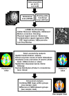
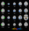
Functional magnetic resonance imaging (fMRI) studies have demonstrated alterations during task-induced brain activation in spinal cord injury (SCI) patients. The interruption to structural integrity of the spinal cord and the resultant disrupted flow of bidirectional communication between the brain and the spinal cord might contribute to the observed dynamic reorganization (neural plasticity). However, the effect of SCI on brain resting-state connectivity patterns remains unclear. We undertook a prospective resting-state fMRI (rs-fMRI) study to explore changes to cortical activation patterns following SCI. With institutional review board approval, rs-fMRI data was obtained in eleven patients with complete cervical SCI (>2 years post injury) and nine age-matched controls. The data was processed using the Analysis of Functional Neuroimages software. Region of interest (ROI) based analysis was performed to study changes in the sensorimotor network using pre- and post-central gyri as seed regions. Two-sampled t-test was carried out to check for significant differences between the two groups. SCI patients showed decreased functional connectivity in motor and sensory cortical regions when compared to controls. The decrease was noted in ipsilateral, contralateral, and interhemispheric regions for left and right precentral ROIs. Additionally, the left postcentral ROI demonstrated increased connectivity with the thalamus bilaterally in SCI patients. Our results suggest that cortical activation patterns in the sensorimotor network undergo dynamic reorganization following SCI. The presence of these changes in chronic spinal cord injury patients is suggestive of the inherent neural plasticity within the central nervous system.
Conflict of interest statement
Figures


Similar articles
-
Evaluation of Whole-Brain Resting-State Functional Connectivity in Spinal Cord Injury: A Large-Scale Network Analysis Using Network-Based Statistic.J Neurotrauma. 2017 Mar 15;34(6):1278-1282. doi: 10.1089/neu.2016.4649. Epub 2017 Jan 27. J Neurotrauma. 2017. PMID: 27937140
-
Alteration of Resting-State Brain Sensorimotor Connectivity following Spinal Cord Injury: A Resting-State Functional Magnetic Resonance Imaging Study.J Neurotrauma. 2015 Sep 15;32(18):1422-7. doi: 10.1089/neu.2014.3661. Epub 2015 Jun 25. J Neurotrauma. 2015. PMID: 25945389
-
Longitudinal evaluation of functional connectivity variation in the monkey sensorimotor network induced by spinal cord injury.Acta Physiol (Oxf). 2016 Jun;217(2):164-73. doi: 10.1111/apha.12645. Epub 2016 Jan 13. Acta Physiol (Oxf). 2016. PMID: 26706280
-
Disentangling the Effects of Spinal Cord Injury and Related Neuropathic Pain on Supraspinal Neuroplasticity: A Systematic Review on Neuroimaging.Front Neurol. 2020 Feb 5;10:1413. doi: 10.3389/fneur.2019.01413. eCollection 2019. Front Neurol. 2020. PMID: 32116986 Free PMC article.
-
Central Nervous System Reorganization and Pain After Spinal Cord Injury: Possible Targets for Physical Therapy-A Systematic Review of Neuroimaging Studies.Phys Ther. 2020 Jun 23;100(6):946-962. doi: 10.1093/ptj/pzaa043. Phys Ther. 2020. PMID: 32201890
Cited by
-
Inconsistency between cortical reorganization and functional connectivity alteration in the sensorimotor cortex following incomplete cervical spinal cord injury.Brain Imaging Behav. 2020 Dec;14(6):2367-2377. doi: 10.1007/s11682-019-00190-9. Brain Imaging Behav. 2020. PMID: 31444779
-
Spinal Cord Injury Disrupts Resting-State Networks in the Human Brain.J Neurotrauma. 2018 Mar 15;35(6):864-873. doi: 10.1089/neu.2017.5212. Epub 2018 Jan 11. J Neurotrauma. 2018. PMID: 29179629 Free PMC article.
-
Brain Network Alterations in Chronic Spinal Cord Injury: Multilayer Community Detection Approach.Neurotrauma Rep. 2024 Nov 6;5(1):1048-1059. doi: 10.1089/neur.2024.0098. eCollection 2024. Neurotrauma Rep. 2024. PMID: 39744611 Free PMC article.
-
Resting-State Functional Magnetic Resonance Imaging Connectivity of the Brain Is Associated with Altered Sensorimotor Function in Patients with Cervical Spondylosis.World Neurosurg. 2018 Nov;119:e740-e749. doi: 10.1016/j.wneu.2018.07.257. Epub 2018 Aug 6. World Neurosurg. 2018. PMID: 30092474 Free PMC article.
-
Integrated Neuroregenerative Techniques for Plasticity of the Injured Spinal Cord.Biomedicines. 2022 Oct 13;10(10):2563. doi: 10.3390/biomedicines10102563. Biomedicines. 2022. PMID: 36289825 Free PMC article.
References
-
- NSCIS. National Spinal Cord Injury Statistical Center Annual Statistical Report for the Spinal Cord Injury Model Systems- Complete Public Version. University of Alabama at Birmingham: Birmingham, Alabama: https://http://www.nscisc.uab.edu. 2015.
-
- Raineteau O, Schwab ME. Plasticity of motor systems after incomplete spinal cord injury. Nature reviews Neuroscience. 2001. April;2(4):263–73. . Epub 2001/04/03. eng. - PubMed
-
- Jurkiewicz MT, Mikulis DJ, McIlroy WE, Fehlings MG, Verrier MC. Sensorimotor Cortical Plasticity During Recovery Following Spinal Cord Injury: A Longitudinal fMRI Study. Neurorehabilitation and Neural Repair. 2007 November 1, 2007;21(6):527–38. - PubMed
-
- Dietz V, Curt A. Neurological aspects of spinal-cord repair: promises and challenges. The Lancet Neurology. 2006. August;5(8):688–94. . Epub 2006/07/22. eng. - PubMed
Publication types
MeSH terms
LinkOut - more resources
Full Text Sources
Other Literature Sources
Medical
Molecular Biology Databases

