

Cochlear changes in serous labyrinthitis associated with silent otitis media: A human temporal bone study
- PMID: 26954857
- PMCID: PMC4785270
- DOI: 10.1016/j.amjoto.2015.10.002
Cochlear changes in serous labyrinthitis associated with silent otitis media: A human temporal bone study
Abstract
Purpose: To determine histopathological findings in the cochlea of human temporal bones with serous labyrinthitis.
Materials and methods: We compared human temporal bones with serous labyrinthitis (20 cases) associated with silent otitis media and without serous labyrinthitis (20 cases) to study location of serous labyrinthitis, the degree of endolymphatic hydrops, number of spiral ganglion cells and hair cells, loss of fibrocytes in the spiral ligament, and areas of the spiral ligament and stria vascularis.
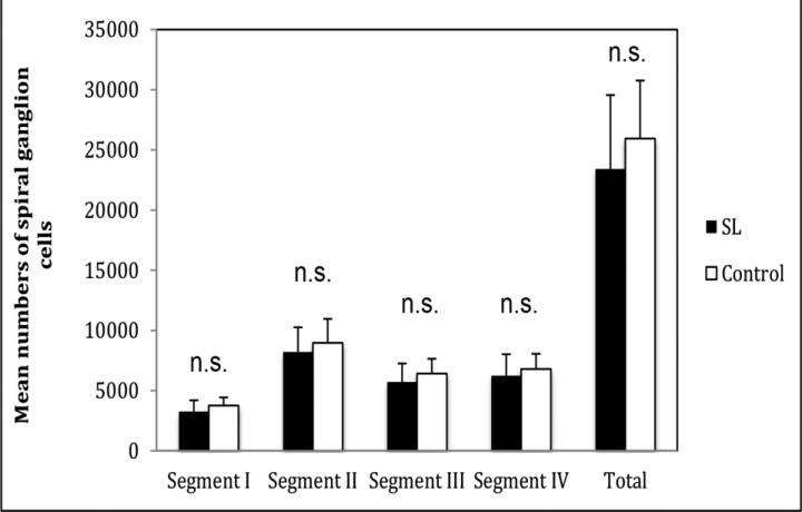
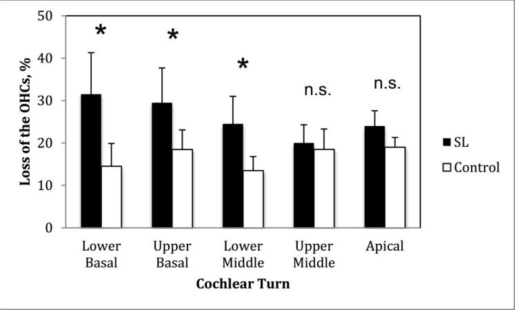
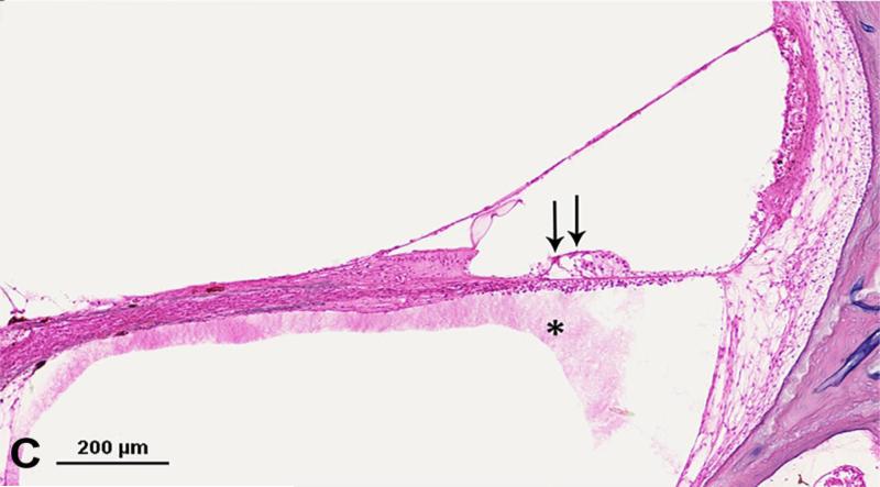
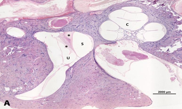
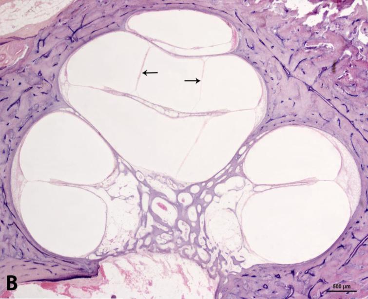
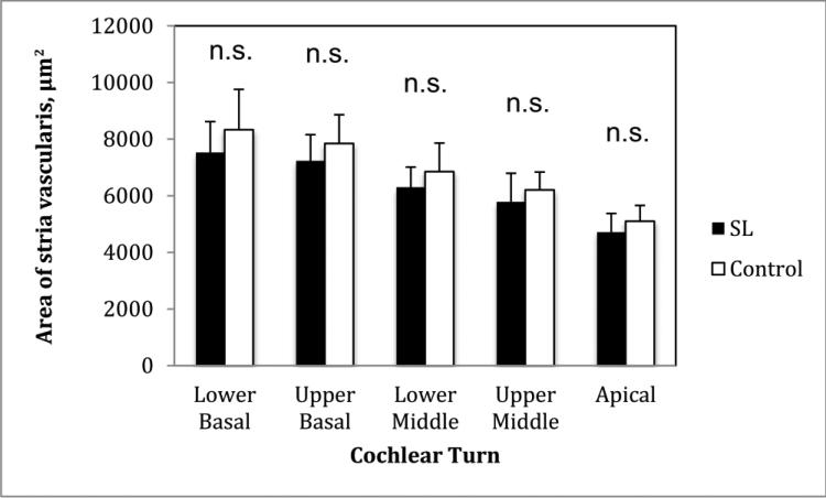
Results: The serous labyrinthitis caused significant loss of outer hair cells in the lower basal (P=0.006), upper basal (P=0.005), and lower middle (P=0.011) cochlear turns, and significant increase in the degree of endolymphatic hydrops than the control group (P=0.036). No significant difference was found in the loss of inner hair cells, in the number of spiral ganglion cells and fibrocytes in the spiral ligament, and in areas of the stria vascularis and spiral ligament (P>0.05).
Conclusions: Serous labyrinthitis resulted in significant loss of outer hair cells and significant increase in the degree of endolymphatic hydrops.
Copyright © 2016 Elsevier Inc. All rights reserved.
Figures
Similar articles
-
Pathologic Findings of the Cochlea in Labyrinthitis Ossificans Associated with the Round Window Membrane.Otolaryngol Head Neck Surg. 2016 Oct;155(4):635-40. doi: 10.1177/0194599816651245. Epub 2016 May 24. Otolaryngol Head Neck Surg. 2016. PMID: 27221575
-
Quantitative Assessment of Cochlear Histopathologic Findings in Patients With Suppurative Labyrinthitis.JAMA Otolaryngol Head Neck Surg. 2016 Apr;142(4):364-9. doi: 10.1001/jamaoto.2015.3803. JAMA Otolaryngol Head Neck Surg. 2016. PMID: 26987015
-
Cochlear changes in chronic otitis media.Laryngoscope. 2004 Apr;114(4):622-6. doi: 10.1097/00005537-200404000-00006. Laryngoscope. 2004. PMID: 15064614
-
Significance of spiral ligament fibrocytes with cochlear inflammation.Int J Pediatr Otorhinolaryngol. 2000 Nov 30;56(1):45-51. doi: 10.1016/s0165-5876(00)00408-0. Int J Pediatr Otorhinolaryngol. 2000. PMID: 11074115 Review.
-
Endolymphatic deafness: a particular variety of cochlear disorder.ORL J Otorhinolaryngol Relat Spec. 2002 Mar-Apr;64(2):120-4. doi: 10.1159/000057790. ORL J Otorhinolaryngol Relat Spec. 2002. PMID: 12021503 Review.
Cited by
-
FGF23 and its role in X-linked hypophosphatemia-related morbidity.Orphanet J Rare Dis. 2019 Feb 26;14(1):58. doi: 10.1186/s13023-019-1014-8. Orphanet J Rare Dis. 2019. PMID: 30808384 Free PMC article. Review.
-
Secondary Endolymphatic Hydrops.Otol Neurotol. 2017 Jun;38(5):774-779. doi: 10.1097/MAO.0000000000001377. Otol Neurotol. 2017. PMID: 28306649 Free PMC article. Review.
-
Relationship Between Cochlear Lateral Wall Changes and Endolymphatic Hydrops in Otitis Media.Laryngoscope. 2024 Dec;134(12):5103-5108. doi: 10.1002/lary.31626. Epub 2024 Jul 3. Laryngoscope. 2024. PMID: 38958129
-
Association Between Acute Otitis Media and Inner Ear Disorders Among Adults in Aseer Region.Cureus. 2021 Nov 14;13(11):e19556. doi: 10.7759/cureus.19556. eCollection 2021 Nov. Cureus. 2021. PMID: 34926038 Free PMC article.
-
Progression of changes in the sensorial elements of the cochlear and peripheral vestibular systems: The otitis media continuum.Hear Res. 2017 Aug;351:2-10. doi: 10.1016/j.heares.2017.05.003. Epub 2017 May 26. Hear Res. 2017. PMID: 28578877 Free PMC article.
References
-
- Cureoglu S, Schachern PA, Rinaldo A, Tsuprun V, Ferlito A, Paparella MM. Round window membrane and labyrinthine pathological changes: an overview. Acta Otolaryngol. 2005;125:9–15. - PubMed
-
- Merchant SN, Nadol JB. Schuknecht's Pathology of the Ear. 3rd ed. People's Medical Publishing House-USA; Shelton, CT: 2010.
-
- Goldstein NA, Casselbrant ML, Bluestone CD, Kurs-Lasky M. Intratemporal complications of acute otitis media in infants and children. Otolaryngol Head Neck Surg. 1998;119:444–54. - PubMed
-
- Arts HA. Sensorineural hearing loss in adults. In: Flint PW, Haughey BH, Lund VJ, et al., editors. Cummings Otolaryngology—Head and Neck Surgery. Saunders-Elsevier; Philadelphia: 2014. pp. 2319–35.
-
- Hellström S, Eriksson PO, Yoon YJ, Johansson U. Interactions between the middle ear and the inner ear: bacterial products. Ann N Y Acad Sci. 1997;830:110–9. - PubMed
Publication types
MeSH terms
Grants and funding
LinkOut - more resources
Full Text Sources
Other Literature Sources
