. 2014 Dec 1;64(12):1073-1083.
doi: 10.1093/biosci/biu180. Epub 2014 Nov 25.

A Structural Basis for How Motile Cilia Beat

Affiliations

A Structural Basis for How Motile Cilia Beat

Peter Satir et al. Bioscience. .

Abstract

The motile cilium is a mechanical wonder, a cellular nanomachine that produces a high-speed beat based on a cycle of bends that move along an axoneme made of 9+2 microtubules. The molecular motors, dyneins, power the ciliary beat. The dyneins are compacted into inner and outer dynein arms, whose activity is highly regulated to produce microtubule sliding and axonemal bending. The switch point hypothesis was developed long ago to account for how sliding in the presence of axonemal radial spoke-central pair interactions causes the ciliary beat. Since then, a new genetic, biochemical, and structural complexity has been discovered, in part, with Chlamydomonas mutants, with high-speed, high-resolution analysis of movement and with cryoelectron tomography. We stand poised on the brink of new discoveries relating to the molecular control of motility that extend and refine our understanding of the basic events underlying the switching of arm activity and of bend formation and propagation.

Keywords: axoneme; cilia; dynein; eukaryotic flagella; microtubules; motility.


Figures

Figure 1.
Figure 1.
Bending patterns of ciliary (a) and flagellar (b) movement in Chlamydomonas captured by high-speed flash photography. The figures illustrate the flexural patterns of movement typical of epithelial cilia and the undulatory patterns of movement typical of eukaryotic flagellar bending. The grey arrows indicate the direction of cell movement. The black bar at the base of the cilia or flagella is 5 micrometers long and marks the site of ciliary or flagellar attachment to the cell. The open arrow in panel (a) indicates the direction of the forward ciliary bend. The open arrow in panel (b) indicates the direction of flagellar bend propagation. Adapted with permission from Brokaw and Kamiya (1987).
Figure 2.
Figure 2.
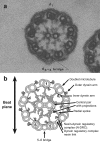
Transverse section of motile cilium. (a) An electron micrograph of a single cilium from the lateral cilia of the clam gill viewed from inside the cell out toward the distal end of the cilium. Doublet 1 is located at the 12 o'clock position, and the other outer doublet microtubules are numbered clockwise in the direction in which the dynein arms point. Also illustrated is the 5–6 bridge. The lateral cilia beat in a plane defined by doublet 1 and the 5–6 bridge, and in these cilia, the effective or forward bend direction is toward the 5–6 bridge. Source: Reprinted with permission from Warner and Satir (1974). (b) Diagram illustrating the main features of the 9 + 2 axoneme from motile metazoan cilia, including the outer doublet and central pair microtubules, the outer and inner dynein arms, the central pair projections, the 5–6 bridge, the radial spokes, and the dynein regulatory complex and nexin links.
Figure 3.
Figure 3.
Diagrams illustrating (a) the phases of ciliary bending and (b) the geometry of a sliding microtubule model for ciliary bending. The details of each diagram are discussed in the text and illustrate the geometry of microtubule sliding for effective and reverse bends for microtubules anchored in the basal body and free to slide at the distal axoneme. The model is based on the discovery that microtubules are inextensible and that the bends are in the form of circular arcs. Source: Reprinted with permission from Satir (1968).
Figure 4.
Figure 4.
The Chlamydomonas axoneme ultrastructure. Cryoelectron tomography slices show (a) a longitudinal, (b) a three-dimensional view, and (c) a cross-sectional view of a Chlamydomonas axoneme. The red boxes highlight one 96-nanometer (nm) axonemal repeat unit in each view. (d, e) Isosurface renderings and (f, g) a simplified schematic show an averaged 96-nm axonemal repeat in (d, f) longitudinal and (e, g) cross-sectional orientation. The cross-sectional slice is taken close to radial spoke 2, viewing from the proximal to the distal end. Key axonemal structures are highlighted: the A- and B-tubules (At, Bt), the nexin-dynein regulatory complex (N-DRC), radial spokes (RS1, RS2), the calmodulin and spoke associated complex (CSC), and the inner and outer dynein arms (IA, OA). The inner arm dyneins include the I1 complex (dynein f α and β) and dyneins a–g. Source: Adapted with permission from Heuser and colleagues (2012a, 2012b).

Similar articles

Cited by

References

    1. Afzelius B. Electron microscopy of the sperm tail; results obtained with a new fixative. Journal of Biophysical and Biochemical Cytology. 1959;5:269–278. - PMC - PubMed
    1. Avasthi P, Marshall WF. Stages of ciliogenesis and regulation of ciliary length. Differentiation. 2012;83:S30–S42. - PMC - PubMed
    1. Barber CF, Heuser T, Carbajal-Gonzalez BI, Botchkarev VV, Jr, Nicastro D. Three-dimensional structure of the radial spokes reveals heterogeneity and interactions with dyneins in Chlamydomonas flagella. Molecular Biology of the Cell. 2012;23:111–120. - PMC - PubMed
    1. Blake JR, Sleigh MA. Mechanics of ciliary locomotion. Biological Reviews of the Cambridge Philosophical Society. 1974;49:85–125. - PubMed
    1. Bower R, Tritschler D, Vanderwaal K, Perrone CA, Mueller J, Fox L, Sale WS, Porter ME. The N-DRC forms a conserved biochemical complex that maintains outer doublet alignment and limits microtubule sliding in motile axonemes. Molecular Biology of the Cell. 2013;24:1134–1152. - PMC - PubMed